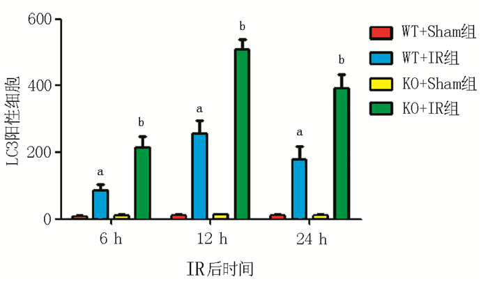
2015 Vol. 6, No. 3

Editorial
2015, 6(3): 131-133. doi: 10.3969/j.issn.1674-7445.2015.03.001
Abstract(70) HTML (52) PDF 120KB($!{article.pdfDownCount})
Abstract:
Expert Forum
2015, 6(3): 134-138. doi: 10.3969/j.issn.1674-7445.2015.03.002
Abstract(97) HTML (39) PDF 207KB($!{article.pdfDownCount})
Abstract:
Original Articles
β-arrestin-2 alleviates mouse hepatic ischemia-reperfusion injury by inhibiting autophagy
Wang Li, Wang Genshu
2015, 6(3): 139-145, 156. doi: 10.3969/j.issn.1674-7445.2015.03.003
Abstract(285) HTML (80) PDF 790KB($!{article.pdfDownCount})
Abstract:
  Objective  To investigate the impact of β-arrestin-2 on hepatic autophagy after ischemia-reperfusion(IR) and its effect on mouse hepatic ischemia-reperfusion injury(IRI).  Methods  β-arrestin-2 wild type(WT) and knock out(KO) mice were used to build a mouse model of hepatic IR(70% hepatic warm ischemia for 90 min). Mice were divided into four groups: WT mice sham-operated group(WT+Sham group), WT mice IR group(WT+IR group), KO mice sham-operated group(KO+Sham group) and KO mice IR group(KO+IR group), 18 mice in each group. Serum and liver tissues were collected at 6, 12 and 24 h after reperfusion. The alanine aminotransferase(ALT), aspartate aminotransferase(AST) measurement and liver tissues hematoxylin-eosin(HE) staining and pathology analysis were used to estimate hepatic injury. The expression of light chain(LC)3, the key protein of autophagy, were detected by immunohistochemical(IHC) staining and western blot. Autophagosomes in liver tissues were evaluated by transmission electron microscopy(TEM).  Results  Compared with Sham groups, the levels of serum ALT and AST significantly increased in IR groups at each time point (all in P < 0.01). The levels of KO+IR group were higher than those of WT+IR group at each time point (all in P < 0.01). The result of liver tissue HE staining showed that liver cell morphology and lobular architecture was normal in WT+Sham group and KO+Sham group at each time point after reperfusion. Liver cells were light or moderate swelling with liver sinus expansion in KO+IR group and WT+IR group at 6 h after reperfusion. And liver cells were severe swelling with inflammatory cells infiltration, and flake damage area is obvious at 12 h after reperfusion. Liver rope arranged regularly at 24 h after reperfusion. The degree of hepatic injury in KO+IR group was more serious than WT+IR group. IHC staining and western blot analysis showed that the expression levels of LC3 increased in IR groups at 6, 12 h but slightly decreased at 24 h after reperfusion. And the expression levels of KO+IR group were higher than WT+IR group. TEM result show that autophagosomes in IR groups were obviously more than those in Sham groups(both in P < 0.01). The counts of autophagosomes in KO+IR group were more than those of WT+IR group(P < 0.05).  Conclusions  β-arrestin-2 may alleviate mouse hepatic IRI by inhibiting autophagy.
Extraction and purification of serum specific endothelial cell antibody of renal transplant recipients
Liu Dan, Hu Juan, Liu Ping, Luo Weiguang, Li Tingting, Guo Jing, Ming Yingzi, Zou Yizhou
2015, 6(3): 146-151. doi: 10.3969/j.issn.1674-7445.2015.03.004
Abstract(109) HTML (65) PDF 326KB($!{article.pdfDownCount})
Abstract:
  Objective  To investigate the extraction and purification methods of serum specific endothelial cell antibody of renal transplant recipients with rejection after renal transplantation.  Methods  Human umbilical vein endothelial cell (HUVEC) was isolated and cultured. The serum samples of the renal transplant recipients with poor renal function after renal transplantation were collected. Specific endothelial cell antibody was screened out with flow cytometry; antibodies against human leukocyte antigen (HLA) and major histocompatibility complex class Ⅰ-related chain A (MICA) were detected by Luminex platform. After the existence of specific endothelial cell antibody in the serum sample was confirmed, specific endothelial cell antibody was absorbed with HUVEC. The cell was washed and then the absorbed antibody was eluted from the cell membrane. Antibody IgG in the eluent was purified and concentrated again with Protein-A/G magnetic beads. Antibody activity in the eluent was detected by flow cytometry and the purified specific endothelial cell antibody (IgG) was identified by SDS-polyacrylamide gel (SDS-PAGE) and Western blot.  Results  In the serum of 386 renal transplant recipients, the serum samples of 5 renal transplant recipients with serum creatinine (Scr) >400 μmoI/L, negative anti-HLA antibody, negative anti-MICA antibody and median fluorescence intensity (MFI) >16 were selected. Purified specific endothelial cell antibody IgG showed immunoglobulin heavy chain (purity >95%) by SDS-PAGE gel. Flow cytometry showed that the purified antibody had the feature of rebinding with the surface antigen of vascular endothelial cell.  Conclusions  The purification method of using human umbilical vein endothelial cell to absorb specific endothelial cell antibody in the serum of renal transplant recipients may obtain good effect.
Effect of GPR49 gene on proliferation and invasive ability of hepatoma cells
Tang Kaiwen, Tang Yunqiang, Gong Yuanfeng, Zhao Hongyu, Wang Jiakang, Tang Hui, Mai Cong
2015, 6(3): 152-156. doi: 10.3969/j.issn.1674-7445.2015.03.005
Abstract(78) HTML (12) PDF 217KB($!{article.pdfDownCount})
Abstract:
  Objective  To discuss the effect of G-protein-coupled receptor 49 (GPR49) gene on proliferation and invasive ability of hepatoma cell line Huh7 and its molecular biological mechanism.  Methods  According to the different transfected small interfering RNA(si-RNA), Huh7 cells were divided into the GPR49-siRNA(si-GPR49)group and the NC-siRNA (si-NC) group. Untransfected Huh7 cells were set as the control group. Messenger RNA(mRNA)and protein expression of GPR49, cyclin D1 and matrix metalloproteinase 9 (MMP9) in the cells of the three groups were respectively detected by reverse transcription-polymerase chain reaction (RT-PCR) and Western blot method. The proliferation and invasive ability of the cells of each group were respectively detected by MTT method and Transwell method.  Results  The relative expression of GPR49 mRNA of the si-GPR49 group was (23.8±3.1)% of the control group (P < 0.05). Compared with the control group, the protein expression of GPR49, cyclin D1 and MMP9 of the si-GPR49 group decreased significantly(all in P < 0.05). The proliferation experiment results by MTT indicated that the optical density(OD)of the cells of the si-GPR49 group at 72 h was (0.53±0.12), which was significantly lower than that of the control group(1.35±0.28). The difference had statistical significance (P < 0.05). The average invaded cell counts of the si-GPR49 group were (13.6±2.5), which was significantly lower than (65.3±6.1) of the control group. The difference had statistical significance (P < 0.05).  Conclusions  GPR49-siRNA may inhibit the gene expression of GPR49 in Huh7 cells. Its mechanism may be that the proliferation of Huh7 cells is inhibited by reducing the level of cyclin D1; the migration and invasive ability of Huh7 cells is inhibited by affecting the expression level of MMP9.
Repairment of 1 cm sciatic nerve defect by bone marrow mesenchymal stem cell of adult rat combined with tissue-engineered artificial nerve
Zhou Lina, Cui Xiaojun, Su Kaixin, Wang Xiaohong, Cai Xueyan, Guo Jinhua, Na Qingqing
2015, 6(3): 157-161, 168. doi: 10.3969/j.issn.1674-7445.2015.03.006
Abstract(95) HTML (39) PDF 351KB($!{article.pdfDownCount})
Abstract:
  Objective  To discuss the effect of bone marrow mesenchymal stem cell (BMSC) as the seed cell transplantation of tissue-engineered artificial nerve in the treatment of peripheral nerve injury.  Methods  BMSC was obtained from the bone marrow of adult rat through isolation and culture and combined with acellular nerve scaffold to construct'tissue-engineered artificial nerve'. After transplantation, rats were divided into two groups, the BMSC+acellular nerve conduit group(BMSC treatment group)and the empty cell conduit group(negative control group)with 5 rats in each group. Sciatic functional index (SFI) of the affected side of rats was compared between two groups at 2 weeks, 4 weeks and 8 weeks after the surgery. Moreover, the sciatic conduction, recovery rate of tricipital muscle wet weight and other repair effects of the affected side were compared between two groups at 8 weeks after the surgery.  Results  The indicators of BMSC treatment group, including SFI assessed at 2 weeks, 4 weeks and 8 weeks after the surgery as well as the sciatic conduction and recovery rate of tricipital muscle wet weight assessed at 8 weeks after the surgery, were better than those of the negative control group(all in P < 0.05).  Conclusions  BMSC combined with tissue-engineered artificial nerve of acellular nerve scaffold can effectively promote nerve regeneration and function recovery.
Protective effect of creatine phosphate on isolated rat liver against cold preservation
Cao Jinglin, Wang Miao, Wang Yang, Ren Guijun, Shi Yanmin, Dou Jian
2015, 6(3): 162-168. doi: 10.3969/j.issn.1674-7445.2015.03.007
Abstract(106) HTML (40) PDF 260KB($!{article.pdfDownCount})
Abstract:
  Objective  To discuss the protective effect of creatine phosphate(CP) on isolated rat liver against cold preservation.  Methods  Isolated perfused rat liver model under simple cold preservation was established. The liver of the control group was perfused with pure University of Wisconsin(UW) solution. With UW solution as the base fluid, the liver of the low-dose group was perfused with 1 g/100 ml CP in UW solution; the liver of the middle-dose group was perfused with 2 g/100 ml CP in UW solution; the liver of the high-dose group was perfused with 3 g/100 ml CP in UW solution. The livers of each group were cold preserved in the corresponding perfusion fluid at 4℃. The content of alanine aminotransferase (ALT) and lactate dehydrogenase (LDH) in preservation solution in infrahepatic vena cava were determined. The content of malondialdehyde (MDA) and activity of myeloperoxidase (MPO) in liver tissues were detected. The apoptosis index (AI) of liver cells in liver tissues and positive expression rate of NF-κB in liver tissues were observed. Pathologic changes of liver tissues were observed under optical microscope.  Results  At 12 h after the cold preservation, the content of ALT and LDH in the rat livers of low-, middle-and high-dose groups were lower than those of the control group (all in P < 0.05). At 18 h after the cold preservation, the content of MDA and MPO in the liver tissues of low-, middle-and high-dose groups were lower than those of the control group (all in P < 0.05). At 12 h and 18 h after the cold preservation, AI and positive expression rate of NF-κB in liver cells in the rat livers of low-, middle-and high-dose groups were lower than those of the control group (all in P < 0.05). At 24 h after the cold preservation, the content of ALT and MDA in preservation solution of the high-dose group was obviously higher than that of the control group as well as the low-and middle-dose groups (all in P < 0.05). The results of pathological examination indicated that the injuries to the livers of the high-, middle-and low-dose groups were obviously lighter than that of the control group. There was no obvious difference among each dose group.  Conclusions  CP in UW solution may well protect the isolated rat liver against cold preservation, which is better than pure UW solution.
Clinical Researches
Trend analysis of malignant tumor after renal transplantation in China
Wang Chao, Li Tao, Zhang Jian, Liu Fen, Xie Zelin, Tang Yawang, Sun Wen, Guo Hongbo, Lin Jun, Zhang Lei, Tian Ye, Ma Linlin
2015, 6(3): 169-173. doi: 10.3969/j.issn.1674-7445.2015.03.008
Abstract(123) HTML (95) PDF 200KB($!{article.pdfDownCount})
Abstract:
  Objective  To reveal the change trend of malignant tumor after renal transplantation in China based on the epidemiological and clinical features that were publicly reported in China in recent 10 years.  Methods  The search terms'renal transplantation' and'tumor'were searched on China Academic Journal Network Publishing Database and China Science Periodical Database to screen out the qualified researches strictly. General conditions, tumor sites and regional differences of malignant tumors were analyzed.  Results  Fifteen thousand one hundred and twenty cases from nine literatures published from 2003 to 2014 and a single-center experience of renal transplantation in Beijing Friendship Hospital of Capital Medical University were screened out. Four hundred and fourty-six cases had malignant tumor after renal transplantation with the total tumor incidence of 2.95% (446/15 120) and the tumors were mainly urinary system tumors after transplantation (55.8%).  Conclusions  The total incidence of malignant tumor in renal transplant recipients is 2.95% and the urinary system tumors are most common. Thus, tumor screening after renal transplantation should be taken as the routine examination during follow-up.
Changes of serum homocysteine level of renal transplant recipients
Kong Xiangrui, Xiao Li, Liu Zhijia, Gao Yu, Bi Lili, Shi Bingyi
2015, 6(3): 174-178. doi: 10.3969/j.issn.1674-7445.2015.03.009
Abstract(102) HTML (56) PDF 228KB($!{article.pdfDownCount})
Abstract:
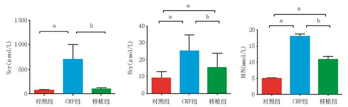
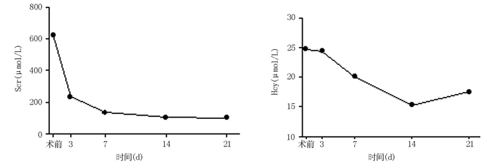
  Objective  To observe the changes of serum homocysteine (Hcy) level of renal transplant recipients before and after renal transplantation, and assess the correlation between serum Hcy level and graft function.  Methods  Thirty-three recipients were included into the transplantation group, who underwent renal allograft transplantation in the Organ Transplant Institute of the 309th Hospital of People's Liberation Army and had renal function recovered stably from January 2013 to June 2014. And 65 patients who were confirmed as chronic renal failure (CRF) by clinical examinations were included into the CRF group and 30 healthy people were included into the control group. A retrospective cross-sectional study was conducted on all of these subjects. Serum Hcy, serum creatinine (Scr) and blood urea nitrogen (BUN) levels of these three groups were compared. Serum Hcy and Scr levels of the transplantation group were continuously monitored before transplantation and at 3, 7, 14 and 21d after transplantation. The correlation between the changes of serum Hcy levels and the renal function before and after transplantation was assessed.  Results  Serum Hcy level of the CRF group was (25±10) μmol/L, which was significantly higher than (9±4) μmol/L of the control group and (15±9) μmol/L of the transplantation group in stable period, with statistical significance (all in P < 0.001). Serum Hcy level of the transplantation group was significantly higher than that of the control group(P < 0.001). Scr level of the CRF group, the transplantation group and the control group was(708±302)μmol/L, (98±23) μmol/L and (72±18) μmol/L, respectively. Scr level of the CRF group was significantly higher than those of the transplantation group and the control group (all in P < 0.001). BUN level of the CRF group, the transplantation group and the control group was (18.1±5.9) mmol/L, (10.9±5.3) mmol/L and (4.9±1.3) mmol/L, respectively. BUN level of the CRF group was significantly higher than that of the transplantation group and the control group (all in P < 0.001), and BUN level of the transplantation group was significantly higher than that of the control group (P < 0.001). With the improvement in renal function after transplantation, Scr and serum Hcy levels of the transplantation group deceased gradually. At 14 d after transplantation, Hcy level decreased to the minimum of (15±5) μmol/L. Compared with (25±10) μmol/L before transplantation, the difference had statistical significance (P < 0.05). Within 14 d after transplantation, serum Hcy level of the transplantation group was positively correlated with Scr level (r=0.761, P < 0.05).  Conclusions  Serum Hcy level of the renal transplant recipients is correlated with the graft function. The combined detection of serum Hcy and renal function index has certain guiding significance in the prevention of hyperhomocysteinemia and the early assessment of graft function.
Analysis on prognosis of pulmonary infection after renal transplantation by Cox risk model
Li Wenhao, Su Zexuan, Ye Zebing, Huang Mingtan, Chen Hang
2015, 6(3): 179-183. doi: 10.3969/j.issn.1674-7445.2015.03.010
Abstract(74) HTML (33) PDF 205KB($!{article.pdfDownCount})
Abstract:
  Objective  To analyze the relevant factors affecting prognosis of pulmonary infection after renal transplantation.   Methods  The clinical data of 40 patients who suffered from pulmonary infection after renal transplantation at the First Affiliated Hospital of Jinan University from January 2000 to December 2010 were analyzed retrospectively. By Cox risk model, single and multi-analysis were performed on 20 possible factors, including age, gender, time of infection, type of infection, white blood cell count, neutrophil ratio, hemoglobin, blood glucose, serum creatinine (Scr), pulmonary infection complicated with acute respiratory distress syndrome or acute pulmonary injury, rejection, blood transfusion, infusion of albumin, infusion of immune globulin, use of ventilator, way of offering oxygen, immunosuppressive regimen, dosage change of immunosuppressant, anti-infection therapeutic regimen and length of stay.   Results and Conclusions  Triple immunosuppressive therapy without mycophenolate mofetil (MMF) and increase of neutrophil ratio were independent risk factors for pulmonary infection after renal transplantation. Triple immunosuppressive therapy with MMF combined with early anti-infection therapeutic regimen may improve patient and graft survival of patients with pulmonary infection after renal transplantation.
Clinical diagnosis and treatment of chronic diarrhea after renal transplantation
Li Zhibin, Zhang Geng, Liu Kepu, Ruan Dongli, Ma Shuaijun, Yang Xiaojian, Qin Weijun, Yuan Jianlin
2015, 6(3): 184-189. doi: 10.3969/j.issn.1674-7445.2015.03.011
Abstract(132) HTML (92) PDF 224KB($!{article.pdfDownCount})
Abstract:
  Objective  To investigate the diagnosis and treatment regimen for patients with chronic diarrhea after renal transplantation.   Methods  The clinical data of 353 patients with chronic diarrhea who underwent renal allograft transplantation at Department of Urology of Xijing Hospital from January 2007 to June 2014 with regular follow-up were analyzed retrospectively. The occurrence of chronic diarrhea after renal transplantation was observed, including incidence, time of occurrence, course of disease and complications. The changes in general conditions and auxiliary examination indexes (body mass index, anemia and other auxiliary examination indexes), treatment and prognosis of the patients with chronic diarrhea were recorded.   Results  Fifteen cases (4.2%)of 353 renal transplant recipients had chronic diarrhea. The time of symptomatic and etiological treatment was (15±7) d. Two patients died during diarrhea (died from gastrointestinal hemorrhage and sudden death caused by severe hypokalemia respectively) and other patients were recovered. Among the 13 patients, 5 cases had good prognosis, 2 cases died(both died from pulmonary infection), 5 cases suffered from renal allograft dysfunction and 1 case suffered from renal allograft insufficiency during the follow-up.   Conclusions  The etiology of chronic diarrhea after renal transplantation is complex and the patients should receive symptomatic and etiological treatment. The patients with chronic diarrhea after renal transplantation combined with severe complications have poor prognosis.
Rh-positive patients receiving kidney grafts from Rh-negative cadaver donor: a report of two cases and literature review
Ma Shuaijun, Zheng Yu, Liu Kepu, Ruan Dongli, Li Zhibin, Zhang Geng, Yang Xiaojian, Yuan Jianlin
2015, 6(3): 190-193. doi: 10.3969/j.issn.1674-7445.2015.03.012
Abstract(106) HTML (52) PDF 176KB($!{article.pdfDownCount})
Abstract:
  Objective  To discuss the safety of Rh-positive patients receiving kidney grafts from Rh-negative cadaver donor.   Methods  On November 29th 2013, two Rh-positive patients received renal transplantation with kidney grafts from Rh-negative cadaver donor at the Department of Urinary Surgery of Xijing Hospital, the Fourth Military Medical University. The clinical data of the two patients were analyzed retrospectively and the relevant literatures were reviewed.   Results  The two patients underwent renal transplantation successfully and no hyperacute rejection or acute rejection occured after the surgery. The two patients were followed up for 12 months. The kidney grafts survived well and the patients had no obvious discomfort.   Conclusions  Through enhanced immunosuppression before operation, sufficient perfusion and close postoperative monitoring, it is safe for Rh-positive patients receiving kidney grafts from Rh-negative cadaver donor.
Hyperthyroidism after allogeneic hematopoietic stem cell transplantation: a case report and literature review
Guo Fang, Zheng Yongjiang
2015, 6(3): 194-196,205. doi: 10.3969/j.issn.1674-7445.2015.03.013
Abstract(128) HTML (138) PDF 175KB($!{article.pdfDownCount})
Abstract:
  Objective  To investigate the characteristics of diagnosis and treatment on hyperthyroidism after allogeneic hematopoietic stem cell transplantation(AHSCT).   Methods  The clinical data of a female patient who suffered from hyperthyroidism after AHSCT at Department of Hematology of the Third Affiliated Hospital, Sun Yat-sen University at December 2008 were analyzed retrospectively. The clinical manifestation, laboratory examination, diagnosis and treatment of the patient were summarized and analyzed, and the relevant literatures were reviewed.   Results  The patient developed the symptoms of hyperthyroidism at 39 months after AHSCT, and had the clinical manifestation and laboratory examination results specific to hyperthyroidism. The patient was diagnosed as diffuse toxic goiter (Graves’ disease) and had good prognosis after drug therapy.   Conclusions  It is rare to develop hyperthyroidism after AHSCT that has specific clinical manifestation and can be confirmed by laboratory examination. Graves' disease has good prognosis with early diagnosis and treatment.
Review Articles
2015, 6(3): 197-200. doi: 10.3969/j.issn.1674-7445.2015.03.014
Abstract(51) HTML (82) PDF 148KB($!{article.pdfDownCount})
Abstract:
2015, 6(3): 201-205. doi: 10.3969/j.issn.1674-7445.2015.03.015
Abstract(88) HTML (33) PDF 193KB($!{article.pdfDownCount})
Abstract:
2015, 6(3): 206-208. doi: 10.3969/j.issn.1674-7445.2015.03.016
Abstract(68) HTML (34) PDF 119KB($!{article.pdfDownCount})
Abstract:
Academic Trend
2015, 6(3): 209-212. doi: 10.3969/j.issn.1674-7445.2015.03.017
Abstract(102) HTML (82) PDF 141KB($!{article.pdfDownCount})
Abstract: