2018 Vol. 9, No. 2

Editorial
2018, 9(2): 91-96. doi: 10.3969/j.issn.1674-7445.2018.01.001
Abstract(429) HTML (262) PDF 1030KB($!{article.pdfDownCount})
Abstract:
Original Articles Experimental Researches
Experimental study on the in vitro induction of regulatory T cells by umbilical cord mesenchymal stem cells with positive human leukocyte antigen-G
Bai Jian, Xiao Li, Miao Lanying, Lin Dayong, Liu Hong, Gao Yu, Chen Wen, Bi Lili, Kong Xiangrui, Huang Haiyan, Shi Bingyi
2018, 9(2): 97-102. doi: 10.3969/j.issn.1674-7445.2018.02.002
Abstract(200) HTML (46) PDF 1365KB($!{article.pdfDownCount})
Abstract:
  Objective  To explore the effect of umbilical cord mesenchymal stem cells with positive human leukocyte antigen (HLA)-G on inducing the production of regulatory T cells (Treg) in vitro.  Methods  Umbilical cord mesenchymal stem cells were isolated from umbilical cord of neonates. PEGFP-N1-HLA-G plasmid was transfected into the human umbilical cord mesenchymal stem cells by liposome transfection, as PEGFP-N1-HLA-G group. PEGFP-N1 empty vector plasmid was transfected into the human umbilical cord mesenchymal stem cells, as PEGFP-N1 group. The human umbilical cord mesenchymal stem cells without empty vector under the same conditions were set as blank control group. Markers of the umbilical cord mesenchymal stem cells were detected using flow cytometry. The expression of HLA-G protein in each group of cells was identified by Western Blot. After mixed-culturing with CD4+T cells in peripheral blood of healthy subjects for 24 h and 48 h, the proportion of CD4+ CD25+ Foxp3+Treg in total T cells of each group was detected by flow cytometry.  Results  CD45, CD34 and HLA-DR presented negative expression on umbilical cord mesenchymal stem cells, while CD29, CD44 and CD105 presented positive expression. HLA-G protein could be expressed in the PEGFP-N1-HLA-G group, which had statistically significant difference compared with the blank control group and PEGFP-N1 group (both P < 0.01). After PEGFP-N1-HLA-G group and CD4+T cells were mixed-cultured for 24 h and 48 h, CD4+ CD25+ Foxp3+Treg accounted for (15.3±1.9)% and (14.3±2.1)% of the total T cells respectively, both of which presented statistically significant difference compared with the blank control group and PEGFP-N1 group (all P < 0.05).  Conclusions  Umbilical cord mesenchymal stem cells with HLA-G gene modified can effectively induce the production of CD4+ CD25+ Foxp3+Treg in vitro.
Effect of human umbilical cord mesenchymal stem cells on CD4+ T cells in liver after hepatic ischemia-reperfusion injury in mice
Zhou Chaorong, Lyu Haijin, Sun Yao, An Yuling, Fan Mingming, Yi Huimin
2018, 9(2): 103-109. doi: 10.3969/j.issn.1674-7445.2018.02.003
Abstract(106) HTML (27) PDF 1646KB($!{article.pdfDownCount})
Abstract:
  Objective  To investigate the effect of human umbilical cord mesenchymal stem cells (HUC-MSCs) on CD4+ T cells in liver after hepatic ischemia-reperfusion injury (HIRI) in mice.  Methods  Two hundred and twentyfive mice were randomly divided into sham group, control group and MSC group, with 75 mice in each group. HIRI model mice were used in MSC group and control group. HUC-MSCs were injected in MSC group through inferior vena cava. Normal saline was injected in control group through inferior vena cava. Only laparotomy and abdominal closure were performed in sham group without blood vessel clipping. At 6, 12 and 24 h after operation, 15 mice of each group were randomly selected to sample eyeball blood and liver tissues, and the 30 mice left in each group were used to extract intrahepatic mononuclear cells. The number of intrahepatic mononuclear cells, percentage, number and positive rate of CD4+ T cells in the mice of various groups at different time points were compared. The content of interleukin (IL)-17 in serum and liver tissue as well as expression levels of costimulatory molecules B7-1 and B7-2 messenger RNA (mRNA) in liver tissues of the mice at different time points were compared.  Results  At 12 and 24 h after operation, the number of intrahepatic mononuclear cells of control group was significantly higher than that of sham group, while the number of intrahepatic mononuclear cells of MSC group was significantly lower than that of control group (P < 0.01-0.05). At 6, 12 and 24 h after operation, the percentage, number and positive rate of CD4+T cells of control group were significantly higher than those of sham group (all P < 0.01), while the percentage of CD4+T cells of MSC group was significantly lower than that of control group (P < 0.01-0.05). At 12 and 24 h after operation, the number and positive rate of CD4+ T cells of MSC group were significantly lower than those of control group (P < 0.01-0.05). At 6, 12 and 24 h after operation, the IL-17 contents in serum and liver tissues of control group were higher than those of sham group (all P < 0.01), while the IL-17 contents in serum and liver tissues of MSC group were lower than those of control group (all P < 0.01). At 6 h after operation, the mRNA expression level of B7-2 of control group was higher than that of sham group (P < 0.05). At 12 and 24 h after operation, the mRNA expression levels of B7-1 and B7-2 of control group were higher than those of sham group (all P < 0.01), while the mRNA expression levels of B7-1 and B7-2 of MSC group were lower than those of control group (all P < 0.01).  Conclusions  HUC-MSCs inhibits the number of CD4+T cells and the secretion of IL-17 in liver after HIRI, as well as decreases the number of intrahepatic mononuclear cells and the mRNA expression of B7-1 and B7-2, thereby alleviating HIRI.
Effect of Nrf2 overexpression on anti-hypoxia and anti-apoptotic ability of mesenchymal stem cells
Liu Rongqiang, Yuan Zenan, Wu Xiaocai, Liu Wei, Wang Guoying
2018, 9(2): 110-115. doi: 10.3969/j.issn.1674-7445.2018.02.004
Abstract(98) HTML (65) PDF 1427KB($!{article.pdfDownCount})
Abstract:
  Objective  To investigate the effect of nuclear factor erythroid-2-related factor 2(Nrf2) on the antihypoxia and anti-apoptotic ability of mesenchymal stem cells(MSCs).  Methods  Human embryonic kidney cells(293FT) were transfected with recombinant plasmid which overexpressed Nrf2 and helper plasmid. High-titer lentivirus which overexpressed Nrf2 were obtained. MSCs were transfected with lentivirus with Nrf2 overexpression and empty lentiviral vector to establish Nrf2-MSCs which stably overexpressed Nrf2 (Nrf2 overexpression group) and green fluorescent protein (GFP)-MSCs(control group). The expression of green fluorescent in 2 groups was observed by fluorescence microscope. The expression level of Nrf2 protein in 2 groups was measured by Western Blot. The anti-hypoxia ability of 2 groups was observed by light microscope. The anti-apoptotic ability of 2 groups was measured by flow cytometry.  Results  Nrf2-MSCs which stably overexpressed Nrf2 were successfully established. Western Blot analysis revealed that the expression level of Nrf2 protein in the Nrf2 overexpression group was significantly higher than that in the control group(P < 0.01). After 15 h hypoxia treatment, the cell activity in the Nrf2 overexpression group was significantly higher than that in the control group. Flow cytometry showed that the apoptosis rate in the Nrf2 overexpression group was (30.9±1.4)%, significantly lower than (61.3±1.3)% in the control group(P < 0.05).  Conclusions  Nrf2-MSCs which can stably overexpress Nrf2 possess certain anti-hypoxia and anti-apoptotic ability in hypoxia environment.
Effect of oxygen glucose deprivation-reperfusion in astrocytes overexpressing endothelin-1 on the proliferation of neural stem/ progenitor cells
Cheng Xiao, Yang Jiajie, Sookjia Kim Chung
2018, 9(2): 116-121. doi: 10.3969/j.issn.1674-7445.2018.02.005
Abstract(155) HTML (74) PDF 1322KB($!{article.pdfDownCount})
Abstract:
  Objective  To investigate the effect of oxygen glucose deprivation-reperfusion (OGD-R) in astrocytes overexpressing endothelin (ET)-1 on the proliferation of neural stem/progenitor cells (NSPCs).  Methods  OGD-R models of negative control astrocytes (C6-Mock) and astrocytes over-expressing ET-1 (C6-ET-1) were constructed. Transwell coculture system of astrocytes and NSPCs was established. Morphologic observation and identification of the astrocytes and primary NSPCs were performed. The cells were divided into four groups: C6-Mock+NSPCs, OGD-R+C6-Mock+NSPCs, C6-ET-1+NSPCs and OGD-R+C6-ET-1+NSPCs groups and co-cultured for 0, 24, 48 and 72 h respectively. The diameter of neurosphere was measured in each group.  Results  In the C6-Mock and C6-ET-1 cells, typeⅠastrocytes in fibrous morphology were observed. Glial fibrillary acidic protein (GFAP) was expressed in the cytoplasm of these two types of cells. Primary NSPCs were positive for nestin staining. After co-culture for 48 and 72 h, the neurosphere diameter in the OGD-R+C6-Mock+NSPCs group was significantly greater than that in the C6-Mock+NSPCs group. The neurosphere diameter in the OGD-R+C6-ET-1+NSPCs group was considerably greater than that in the C6-ET-1+NSPCs group. The neurosphere diameter in the OGD-R+C6-ET-1+NSPCs group was significantly greater compared with that in the OGD-R+C6-Mock+NSPCs group (all P < 0.05).  Conclusions  OGD-R astrocytes can promote the proliferation of NSPCs. ET-1 over-expression further accelerates the proliferation of NSPCs.
Effect of high expression of Zwint on the proliferation of hepatoma cells and the prognosis of liver transplantation for hepatocellular carcinoma
Li Hui, Wang Genshu, Zheng Jun, Cai Jianye, Zhang Junbin, Cheng Daorou, Zhou Qi, Yang Yang
2018, 9(2): 122-129. doi: 10.3969/j.issn.1674-7445.2018.02.006
Abstract(144) HTML (50) PDF 1635KB($!{article.pdfDownCount})
Abstract:
  Objective  To investigate the expression of zeste white 10 interactor (Zwint) in primary hepatocellular carcinoma (HCC) and its effect on the prognosis of liver transplantation for HCC.  Methods  HCC tissues, paracancerous tissues and clinical data of 50 liver transplant recipients for HCC were collected. The expression levels of Zwint messenger RNA (mRNA) and Zwint protein in 20 pairs of HCC tissues and paracancerous tissues of 20 liver transplant recipients for HCC were compared using real-time fluorescence quantitative polymerase chain reaction (PCR), Western Blot and immunohistochemistry (IHC). Two HCC cell lines HepG-2 which interfered with the expression of Zwint successfully were selected as si-Zwint-1 group and si-Zwint-2 group, and the blank control was taken as si-NC group. The cell proliferation and cell cycle of various groups were compared using cell counting kit (CCK) -8 experiment, flatcloning assay and cell cycle experiment. The consistency of the expression of Zwint and cyclin D1 in HCC tissues and cells was analyzed using Western Blot and IHC. The enrolled patients were divided into high expression group (22 cases) and low expression group (28 cases) based on the median of Zwint protein expression level, and the relationship of the expression level of Zwint protein and clinical characteristics, overall survival rate and disease free survival rate of liver transplant recipients for HCC was analyzed.  Results  The results of real-time fluorescence quantitative PCR showed that the expression level of Zwint mRNA in HCC tissues was higher than that of paracancerous tissues (P=0.03). The results of Western Blot and IHC showed that the expression level of Zwint protein in HCC tissues was higher than that of paracancerous tissues (both P < 0.05). After the Zwint gene of HCC cell line HepG-2 was interfered, CCK-8 and flatcloning assay showed that the cell proliferation potential was significantly weakened (all P < 0.01), and the cell cycle arrested at stage G1 (all P < 0.05). The expression level of Zwint protein was closely related to tumor diameter and tumor, node, metastasis (TNM) staging (all P < 0.05). The overall survival rate of liver transplant recipients for HCC in the high Zwint expression group was lower than that of the low expression group (P=0.02).  Conclusions  Zwint is highly expressed in HCC tissues, and it can promote the proliferation of HCC cells through regulating cell cycle. The expression level of Zwint is negatively correlated with the prognosis of liver transplantation for HCC.
Original Articles Clinical Researches
Analysis of risk factors of early acute kidney injury after liver transplantation from DCD donor liver
Wang Wanli, Li Qingshan, Zhou Ying, Wang Li, Sha Huanchen, Tian Min, Shi Jianhua, Dong Jian, Liu Xuemin, Zhang Xiaogang, Liu Chang, Yu Liang, Lyu Yi, Wang Bo
2018, 9(2): 130-136. doi: 10.3969/j.issn.1674-7445.2018.02.007
Abstract(111) HTML (73) PDF 1119KB($!{article.pdfDownCount})
Abstract:
  Objective  To analyze the risk factors of early acute kidney injury (AKI) after liver transplantation from donation after cardiac death(DCD) donor liver.  Methods  Clinical data of 184 donors and recipients undergoing liver transplantation from DCD donor liver were retrospectively analyzed. According to the incidence of early AKI, all participants were divided into the AKI and non-AKI groups. The patients in the AKI group were subject to AKI staging. Baseline data, preoperative, intraoperative and postoperative related parameters were statistically compared between two groups. The cumulative survival rate and clinical prognosis of patients in non-AKI group and AKI group with different staging were statistically analyzed by Kaplan-Meier curve analysis.  Results  Among 184 patients, 68 cases (37.0%) presented with early AKI after liver transplantation including 31 stage 1 AKI, 26 stage 2 AKI and 11 stage 3 AKI, mainly occurring within postoperative 3 d. Univariate analysis revealed that preoperative levels of albumin < 35 g/L, preoperative levels of serum sodium ≤137 mmol/L, operation time>7.5 h, intraoperative hemorrhage volume>3 000 mL, intraoperative red cell infusion volume>15 U and intraoperative urine amount ≤100 mL/h were the risk factors of early AKI after liver transplantation (all P < 0.05). Multi-variate Logistic regression analysis demonstrated that intraoperative red cell infusion > 15 U was an independent risk factor of early AKI after liver transplantation [odds ratio(OR) 1.061, 95% confidence interval(CI) 1.008-1.118, P=0.024]. Result of Kaplan-Meier survival curve suggested that the cumulative survival rate was gradually reduced along with the aggravation of AKI with statistical significance (all P < 0.05).  Conclusions  The incidence of early AKI following liver transplantation is relatively high. The severity of early AKI is intimately correlated with the short-and long-term prognosis of the recipients. A large quantity of intraoperative red blood cell infusion is an independent risk factor of AKI.
Diagnostic value of flow cytometry in postoperative infection after renal transplantation
Ma Xihui, Gao Yu, Han Yong, Sun Yujie, Du Ruo, Liu Peixia, Zhang Wenhui, Xiao Li
2018, 9(2): 137-141,155. doi: 10.3969/j.issn.1674-7445.2018.02.008
Abstract(189) HTML (56) PDF 1178KB($!{article.pdfDownCount})
Abstract:
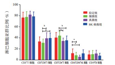
  Objective  To assess the value of flow cytometry in the diagnosis of postoperative infection following renal transplantation.  Methods  According to postoperative imaging findings and laboratory examination outcomes, 51 recipients undergoing the first renal transplantation were divided into the bacteria (n=33), fungus (n=9) and BK virus (n=9) groups. Twenty-eight recipients with stable conditions after renal transplantation were assigned into the stable group. Flow cytometry was adopted to detect the percentage and absolute counting of lymphocyte subpopulation in the peripheral blood of recipients in each group. Renal function, percentage and absolute counting of lymphocyte subpopulation in the peripheral blood were statistically compared among different groups. Receiver operating characteristic (ROC) curve was drawn to evaluate the diagnostic value of the percentage and absolute counting of lymphocyte subpopulation in infectious diseases after renal transplantation.  Results  Compared with the stable group, the serum creatinine (Scr) and blood urea nitrogen (BUN) levels in the bacteria, fungus and BK virus groups were significantly up-regulated, respectively (P=0.035, 0.007, 0.024; 0.037, 0.006, 0.032). Compared with the stable group, the percentage of CD16+ CD56+ natural killer (NK) cells was significantly declined in the bacterial (P=0.036) and fungus groups (P=0.015), and the proportion of CD4+ /CD8+T cells was dramatically decreased in the fungus group (P=0.004). Compared with the bacterial group, the percentage of CD3+ CD8+T cells was significantly elevated (P=0.013 and 0.008), the proportion of CD3+ CD4+T cells was considerably declined (P=0.003 and 0.010), and the percentage of CD4+/CD8+T cells was significantly declined (P=0.003 and 0.005) in the fungus and BK virus groups. Compared with the stable group, the quantity of CD3+ T cells, CD3+ CD8+T cells and CD16+ CD56+ NK cells was significantly declined in the bacterial, fungus and BK virus groups, respectively (P=0.025, 0.002, 0.003; 0.015, 0.005, 0.006; 0.001, 0.001, 0.031). In addition, the quantity of CD3+ CD4+T cells was considerably decreased in the fungus and BK virus groups (P=0.001, 0.003). The quantity of CD19+ B cells was significantly reduced in the BK virus group (P=0.019). Compared with the bacterial group, the quantity of CD3+ CD4+T cells was considerably lower in the fungus group (P=0.023). ROC curve analysis revealed that the quantity of CD3+ CD4+T cells [area under curve(AUC)=0.8492] and CD16+ CD56+ NK cells (AUC=0.8889) yielded relatively high accuracy in the diagnosis of fungal infection. The quantity of CD3+ T cells (AUC=0.8472), CD3+ CD4+T cells (AUC=0.8452) and CD19+ B cells (AUC=0.8115) yielded relatively high accuracy in the diagnosis of BK virus infection.  Conclusions  Flow cytometry detection of the lymphocyte subpopulation in peripheral blood can evaluate the immune function of patients. Absolute counting of lymphocyte subpopulation can directly assess the degree of immunity. These two combined parameters provide guiding significance for the diagnosis and differential diagnosis of infectious diseases in recipients after renal transplantation.
Analysis of risk factors for cerebral apoplexy in the recipients after renal transplantation
Du Peng, Zhang Pengjie, Duan Bin, Chen Rui, Ding Tong
2018, 9(2): 142-146. doi: 10.3969/j.issn.1674-7445.2018.02.009
Abstract(153) HTML (39) PDF 1044KB($!{article.pdfDownCount})
Abstract:
  Objective  To analyze the risk factors for cerebral apoplexy in the recipients after renal transplantation.  Methods  Clinical data of 376 renal transplant recipients who were followed up regularly were retrospectively analyzed. The recipients were divided into cerebral apoplexy group (39 cases) and non-cerebral apoplexy group (337 cases) according to the occurrence of cerebral apoplexy. The risk factors of cerebral apoplexy were analyzed using single factor analysis and COX proportional hazards regression model.  Results  The 376 recipients were followed up for a median duration of 55 months, among whom 39 recipients suffered from cerebral apoplexy, with a cumulative incidence of 10.4%. Single factor analysis indicated that there were significant differences in age ≥40 years old at transplantation, duration of dialysis ≥12 months before transplantation, estimated glomerular filtration rate (eGFR) < 30 mL/(min·1.73m2), incidence of hypertension, diabetes and dyslipidemia between cerebral apoplexy group and non-cerebral apoplexy group (all P < 0.05). Multivariate analysis indicated that the independent risk factors for cerebral apoplexy occurred in the recipients after renal transplantation were age ≥40 years old [hazard ratio (HR) =1.110, 95% confidence interval (CI)=1.067-1.154, P=0.000], duration of dialysis ≥12 months before transplantation (HR=1.044, 95%CI=1.021-1.067, P=0.000) and eGFR < 30 mL/(min·1.73m2) (HR=2.448, 95%CI=1.197-5.005, P=0.014).  Conclusions  The independent risk factors for cerebral apoplexy in the recipients after renal transplantation include age ≥ 40 years old, long duration of dialysis before transplantation and renal insufficiency.
Characteristics and risk factors analysis of infection after liver transplantation from donor liver of donation after citizen's death
Gong Xueyi, Luo Qijie, He Kun, Hu Zemin
2018, 9(2): 147-151. doi: 10.3969/j.issn.1674-7445.2018.02.010
Abstract(163) HTML (82) PDF 1028KB($!{article.pdfDownCount})
Abstract:
  Objective  To investigate the characteristics and risk factors of infection after liver transplantation from donor liver of donation after citizen's death.  Methods  Clinical data of 68 recipients after liver transplantation from donor liver of donation after citizen's death were analyzed retrospectively. The recipients were divided into infection group (33 cases) and non-infection group (35 cases) according to the presence of infection after operation. Major infection characteristics of the 68 recipients after liver transplantation were summarized. Univariate analysis was conducted on the possible risk factors of infection after liver transplantation, and multivariate analysis was further conducted on the risk factors with statistical significance, so as to find out the independent risk factors. In addition, accuracy of predicting infection after liver transplantation was analyzed using receiver operating characteristic (ROC) curves.  Results  Thirtythree recipients were infected after liver transplantation, accounting for 49% of the total recipients with bacterial infection and fungal infection mainly. These recipients mainly presented pulmonary infection and abdominal cavity infection. Univariate analysis results showed that a total of 8 factors contributed to infection after liver transplantation from donor liver of organ donation, including the donors' open injury, recipients' preoperative hemoglobin level, platelet count, ChildPugh classification of liver function, model for end-stage liver disease (MELD) score, intraoperative erythrocyte infusion, gamma-glutamyl transpeptidase (GGT) on day 1 after operation and postoperative stay time of intensive care unit (ICU) (all P < 0.05). Multivariate Logistic regression results analysis showed that preoperative hemoglobin level < 120 g/L and postoperative stay time of ICU >96 h were the independent risk factors of infection after liver transplantation from donor liver of organ donation (both P < 0.05). Analysis results of ROC curves showed that preoperative hemoglobin level < 114 g/L and postoperative stay time of ICU >102 h resulted in higher accuracy for predicting postoperative infection.  Conclusions  Infection after liver transplantation from donation after citizen's death presents high incidence, dominated by bacterial infection and fungal infection in lung and abdominal cavity. Low preoperative hemoglobin level and long postoperative stay time of ICU of recipients can increase the risk of infection after liver transplantation.
Short Article
2018, 9(2): 152-155. doi: 10.3969/j.issn.1674-7445.2018.02.011
Abstract(205) HTML (75) PDF 939KB($!{article.pdfDownCount})
Abstract:
Review Articles
2018, 9(2): 156-158. doi: 10.3969/j.issn.1674-7445.2018.02.012
Abstract(161) HTML (71) PDF 866KB($!{article.pdfDownCount})
Abstract:
2018, 9(2): 159-161, 168. doi: 10.3969/j.issn.1674-7445.2018.02.013
Abstract(107) HTML (38) PDF 942KB($!{article.pdfDownCount})
Abstract:
2018, 9(2): 162-165. doi: 10.3969/j.issn.1674-7445.2018.02.014
Abstract(161) HTML (62) PDF 938KB($!{article.pdfDownCount})
Abstract:
2018, 9(2): 166-168. doi: 10.3969/j.issn.1674-7445.2018.02.015
Abstract(184) HTML (78) PDF 885KB($!{article.pdfDownCount})
Abstract: