Analysis of three-dimensional visualization imaging of severe portal vein stenosis after liver transplantation and clinical efficacy of portal vein stent implantation
-
摘要:
目的 分析肝移植术后严重门静脉狭窄的三维成像特征与优势,评估门静脉支架植入术效果。 方法 回顾性分析10例肝移植术后因严重门静脉狭窄接受门静脉支架植入的患者的临床资料,分析严重门静脉狭窄的影像学特征、三维重建的成像优势及介入治疗效果。 结果 10例患者中狭窄类型包括向心性缩窄3例,曲折成角致狭窄2例,受压狭窄2例,长段狭窄和(或)血管闭塞3例。三维重建图像在狭窄的准确判断、狭窄类型的辨别和狭窄累及长度判断方面具有优势。所有患者均成功接受门静脉支架植入术,支架植入后门静脉最狭窄处直径较治疗前增加[(6.2±0.9)mm比(2.6±1.7)mm,P<0.05],吻合口流速较治疗前下降[(57±19)cm/s比(128±27)cm/s,P<0.05],近肝处门静脉主干流速较治疗前增加[(41±6)cm/s比(18±6)cm/s,P<0.05]。1例患者因介入穿刺引起肝内血肿,经保守观察治疗后好转,其余患者均未出现相关并发症。 结论 三维可视化技术可以立体直观展示狭窄部位、特征与严重程度,有利于临床医师进行治疗决策和辅助介入操作。及时的门静脉支架植入术可以有效逆转病变进程并改善门静脉血流。 Abstract:Objective To analyze three-dimensional imaging characteristics and advantages for severe portal vein stenosis after liver transplantation, and to evaluate clinical efficacy of portal vein stent implantation. Methods Clinical data of 10 patients who received portal vein stent implantation for severe portal vein stenosis after liver transplantation were retrospectively analyzed. Imaging characteristics of severe portal vein stenosis, and advantages of three-dimensional reconstruction imaging and interventional treatment efficacy for severe portal vein stenosis were analyzed. Results Among 10 patients, 3 cases were diagnosed with centripetal stenosis, tortuosity angulation-induced stenosis in 2 cases, compression-induced stenosis in 2 cases, long-segment stenosis and/or vascular occlusion in 3 cases. Three-dimensional reconstruction images possessed advantages in accurate identification of stenosis, identification of stenosis types and measurement of stenosis length. All patients were successfully implanted with portal vein stents. After stent implantation, the diameter of the minimum diameter of portal vein was increased [(6.2±0.9) mm vs. (2.6±1.7) mm, P<0.05], the flow velocity at anastomotic site was decreased [(57±19) cm/s vs. (128±27) cm/s, P<0.05], and the flow velocity at the portal vein adjacent to the liver was increased [(41±6) cm/s vs. (18±6) cm/s, P<0.05]. One patient suffered from intrahepatic hematoma caused by interventional puncture, which was mitigated after conservative observation and treatment. The remaining patients did not experience relevant complications. Conclusions Three-dimensional visualization technique may visually display the location, characteristics and severity of stenosis, which is beneficial for clinicians to make treatment decisions and assist interventional procedures. Timely implantation of portal vein stent may effectively reverse pathological process and improve portal vein blood flow. -

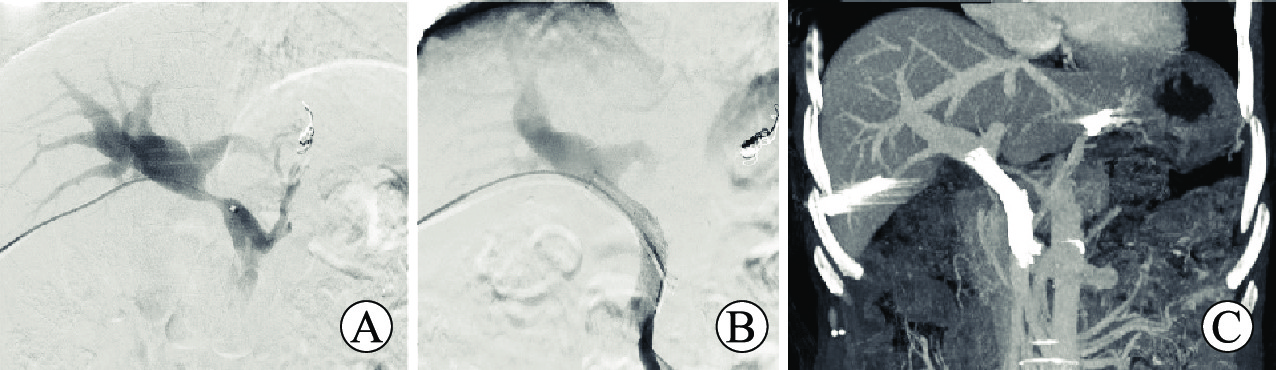
图 1 经皮经肝穿刺介入支架植入治疗肝移植术后门静脉狭窄
注:A图为门静脉造影,显示狭窄区域;B图为支架植入并造影显示狭窄改善情况;C图为术后CT提示门静脉血流通畅,血管直径正常。
Figure 1. Percutaneous transhepatic puncture and stent implantation for the treatment of portal vein stenosis after liver transplantation

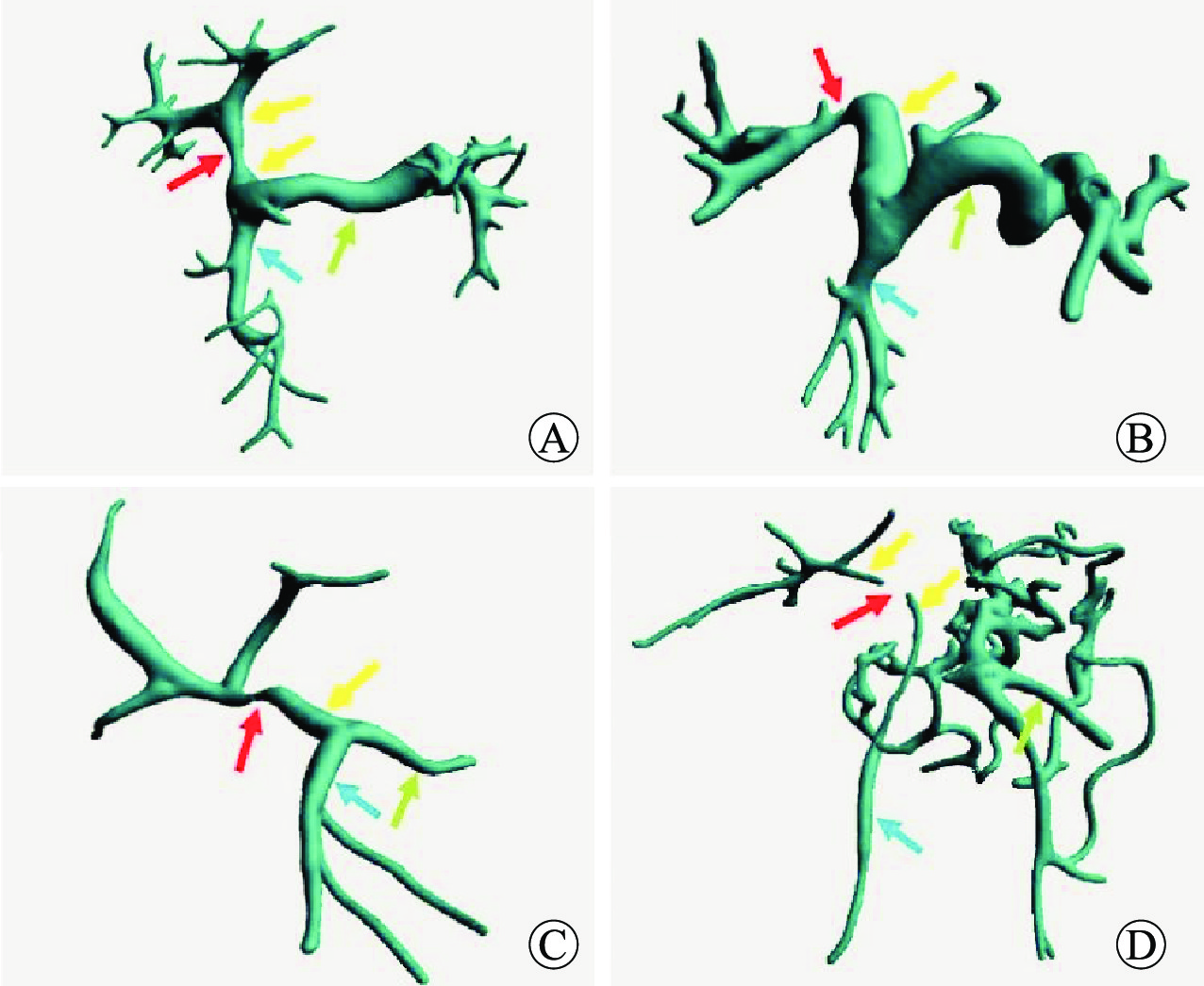
图 2 三维重建显示肝移植术后门静脉、肠系膜上静脉、脾静脉重建图像及相应狭窄情况
注:A图示向心性缩窄,血管环周均匀向中心内缩直径变小(红色箭头所示);B图示曲折成角致狭窄,亲属活体右半肝肝移植术后门静脉右支与门静脉主干之间成角(红色箭头所示)导致狭窄;C图示受压狭窄,可见门静脉受压(红色箭头所示)偏心性狭窄并向腹侧推挤;D图示长段狭窄并伴随区域血管闭塞,可见门静脉长距离纤细,且局部结构消失(红色箭头所示)。黄色箭头示门静脉,蓝色箭头示肠系膜上静脉,绿色箭头示脾静脉。
Figure 2. 3D reconstruction images of portal vein, superior mesenteric vein, splenic vein and corresponding stenosis after liver transplantation
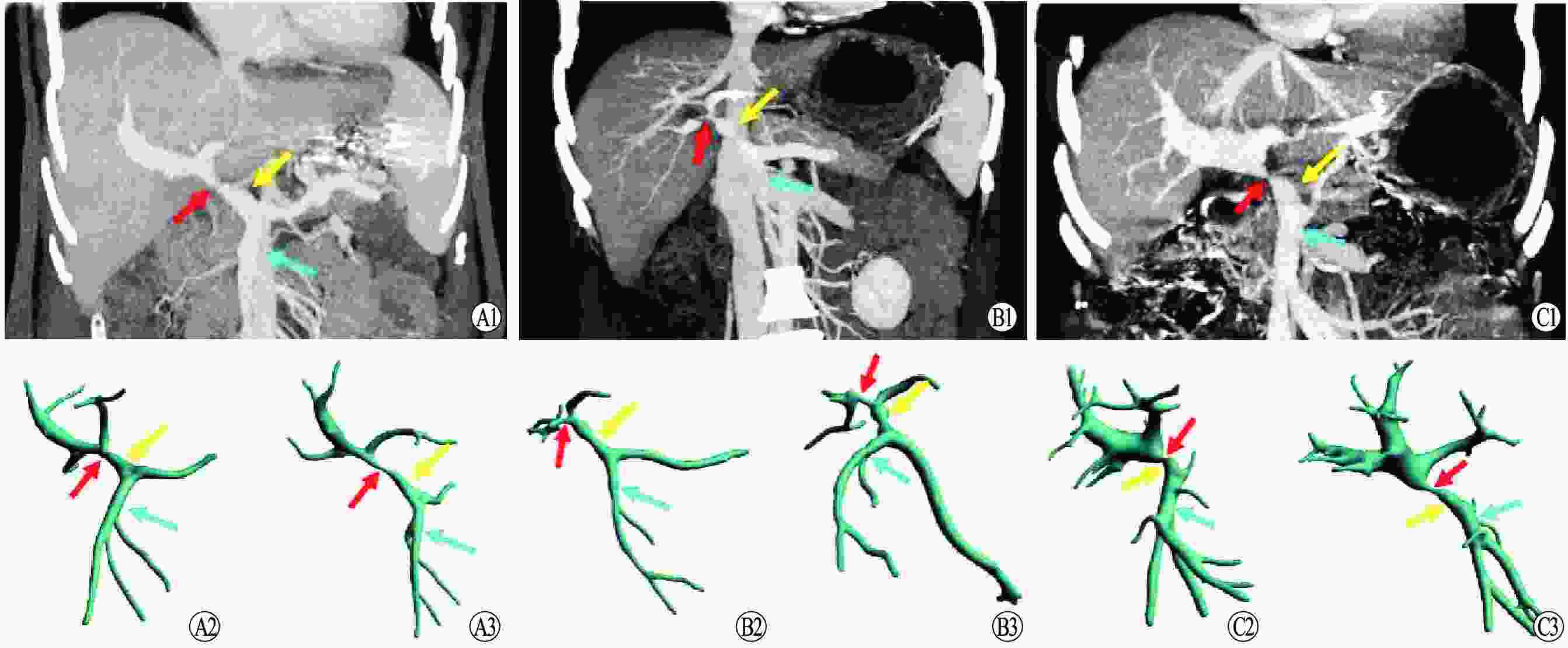
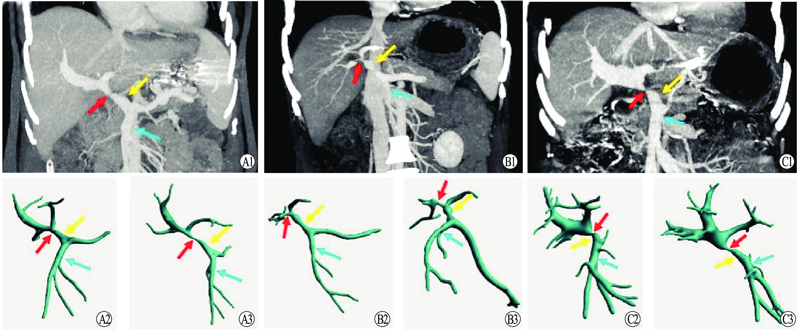
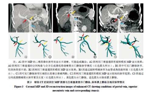
图 3 增强CT的冠状位MIP图像与三维重建显示门静脉、肠系膜上静脉及相应狭窄情况
注:A1图中MIP的二维图像在狭窄处显示不清晰,可能造成漏诊;A2图利用三维重建图形旋转模拟MIP显示效果;A3图利用三维重建的空间构象与全方位成像优势清晰展示门静脉狭窄情况(红色箭头所示);B1图中可见门静脉狭窄,但具体类型不清;B2图利用三维重建图形模拟MIP显示效果;B3图通过旋转明确狭窄为血管成角扭曲所致(红色箭头所示);C1图可见门静脉狭窄区域但长度难以准确判断;C2图利用三维重建图形模拟MIP显示较短的狭窄范围;C3图通过空间成像精确展示狭窄累及长度(红色箭头所示)。黄色箭头示门静脉,蓝色箭头示肠系膜上静脉。
Figure 3. Coronal MIP and 3D reconstruction images of enhanced CT showing conditions of portal vein, superior mesenteric vein and corresponding stenosis
表 1 10例肝移植术后门静脉狭窄接受介入治疗患者的临床资料
Table 1. Clinical data of 10 patients with portal vein stenosis undergoing interventional treatment after liver transplantation
例序 性别 年龄
(岁)原发病 门静脉
情况血栓分级 手术方式 术后狭窄发生
时间(d)狭窄
类型支架相关
并发症随访时间(月) 远期支架
再狭窄1 女 38 肝包虫病 包虫压迫闭塞 Yerdel Ⅱ级 经典原位 204 中心缩窄 无 50 无 2 男 54 原发性肝细胞癌 门静脉海绵样变性 Yerdel Ⅱ级 改良背驮 512 长段狭窄闭塞 无 22 无 3 男 42 不明原因肝硬化 门静脉海绵样变性 Yerdel Ⅱ级 经典原位 6 长段狭窄闭塞 无 15 无 4 男 42 原发性肝细胞癌 门静脉
血栓Yerdel Ⅰ级 改良背驮 113 中心缩窄 无 19 无 5 男 37 乙肝肝硬化 门静脉海绵样变性 Yerdel Ⅲ级 改良背驮 3 长段狭窄 无 12 无 6 男 30 乙肝肝硬化 正常 无 亲属活体右半肝 194 曲折成角 肝内血肿 50 无 7 男 51 原发性肝细胞癌 灭活癌栓 Yerdel Ⅱ级 经典原位 112 中心缩窄 无 40 无 8 女 50 原发性肝细胞癌 TIPS①支架术后 Yerdel Ⅰ级 经典原位 61 挤压狭窄 无 10 无 9 女 51 原发性胆汁性肝硬化 TIPS支架术后 Yerdel Ⅰ级 经典原位 159 挤压狭窄 无 2 无 10 女 38 血管周围上皮样细胞肿瘤 受压变形 无 自体肝
移植515 曲折成角 无 16 无 注:①TIPS为经颈静脉肝内门体静脉分流术。  下载: 导出CSV
表 2 10例肝移植术后门静脉狭窄患者介入治疗前后指标变化[M(P25,P75)]
Table 2. The index changes of 10 patients with portal vein stenosis after liver transplantation before and after interventional therapy
时间 n ALT(U/L) AST(U/L) TB(μmol/L) ALP(U/L) 白细胞(×109/L) 血小板(×109/L) 治疗前 10 34(24, 84) 39(29, 51) 12(10, 30) 120(89, 139) 2.8(2.7, 3.7) 118(38, 164) 治疗后 10 26(19, 57) 23(19, 26) 12(10, 21) 104(91, 126) 4.1(3.0, 4.8) 155(117, 168) P值 0.353 0.052 0.684 0.631 0.075 0.247
下载: 导出CSV
-
[1] BUROS C, DAVE AA, FURLAN A. Immediate and late complications after liver transplantation[J]. Radiol Clin North Am, 2023, 61(5): 785-795. DOI: 10.1016/j.rcl.2023.04.002. [2] KIM KS, KIM JM, LEE JS, et al. Stent insertion and balloon angioplasty for portal vein stenosis after liver transplantation: long-term follow-up results[J]. Diagn Interv Radiol, 2019, 25(3): 231-237. DOI: 10.5152/dir.2019.18155. [3] BUKOVA M, FUNKEN D, PFISTER ED, et al. Long-term outcome of primary percutaneous stent angioplasty for pediatric posttransplantation portal vein stenosis[J]. Liver Transpl, 2022, 28(9): 1463-1474. DOI: 10.1002/lt.26488. [4] MULLAN CP, SIEWERT B, KANE RA, et al. Can Doppler sonography discern between hemodynamically significant and insignificant portal vein stenosis after adult liver transplantation?[J]. AJR Am J Roentgenol, 2010, 195(6): 1438-1443. DOI: 10.2214/AJR.10.4636. [5] YABUTA M, SHIBATA T, SHIBATA T, et al. Long-term outcome of percutaneous transhepatic balloon angioplasty for portal vein stenosis after pediatric living donor liver transplantation: a single institute's experience[J]. J Vasc Interv Radiol, 2014, 25(9): 1406-1412. DOI: 10.1016/j.jvir.2014.03.034. [6] 魏欣, 胡鸿, 钟立明, 等. 术前模拟在经颈静脉肝内门体静脉分流术治疗中的价值[J]. 中国普外基础与临床杂志, 2019, 26(12): 1424-1428. DOI: 10.7507/1007-9424.201908053.WEI X, HU H, ZHONG LM, et al. Value of preoperative simulation in the treatment of transjugular intrahepatic portosystemic shunt[J]. Chin J Bases Clin Gen Surg, 2019, 26(12): 1424-1428. DOI: 10.7507/1007-9424.201908053. [7] SCHOENHAGEN P, NUMBURI U, HALLIBURTON SS, et al. Three-dimensional imaging in the context of minimally invasive and transcatheter cardiovascular interventions using multi-detector computed tomography: from pre-operative planning to intra-operative guidance[J]. Eur Heart J, 2010, 31(22): 2727-2740. DOI: 10.1093/eurheartj/ehq302. [8] DE ASSIS A, DE CARVALHO MELO JA JR, KAWAKAMI WY, et al. Life-threatening variceal bleeding after liver transplantation and extensive portal vein thrombosis: desperate times call for desperate measures[J]. Pediatr Transplant, 2023, 27(8): e14555. DOI: 10.1111/petr.14555. [9] DUFFY JP, HONG JC, FARMER DG, et al. Vascular complications of orthotopic liver transplantation: experience in more than 4, 200 patients[J]. J Am Coll Surg, 2009, 208(5): 896-903; discussion 903-905. DOI: 10.1016/j.jamcollsurg.2008.12.032. [10] 贺英杰, 温培豪, 张嘉凯, 等. 肝移植术后早期门静脉并发症的危险因素分析及诊治经验[J]. 中华器官移植杂志, 2019, 40(11): 660-664. DOI: 10.3760/cma.j.issn.0254-1785.2019.11.003.HE YJ, WEN PH, ZHANG JK, et al. Risk factor analysis and diagnosis and treatment of early portal vein complications after liver transplantation[J]. Chin J Organ Transplant, 2019, 40(11): 660-664. DOI: 10.3760/cma.j.issn.0254-1785.2019.11.003. [11] HU LS, ZHAO Z, LI T, et al. The management of portal vein thrombosis after adult liver transplantation: a case series and review of the literature[J]. J Clin Med, 2022, 11(16): 4909. DOI: 10.3390/jcm11164909. [12] 张佳斌, 栗光明. 成人肝移植术后血管并发症的诊治[J]. 器官移植, 2022, 13(5): 555-560. DOI: 10.3969/j.issn.1674-7445.2022.05.002.ZHANG JB, LI GM. Diagnosis and treatment of vascular complications after liver transplantation in adults[J]. Organ Transplant, 2022, 13(5): 555-560. DOI: 10.3969/j.issn.1674-7445.2022.05.002. [13] NACIF LS, ZANINI LY, PINHEIRO RS, et al. Portal vein surgical treatment on non-tumoral portal vein thrombosis in liver transplantation: systematic review and meta-analysis[J]. Clinics (Sao Paulo), 2021, 76: e2184. DOI: 10.6061/clinics/2021/e2184. [14] MARRA P, DULCETTA L, CARBONE FS, et al. The role of imaging in portal vein thrombosis: from the diagnosis to the interventional radiological management[J]. Diagnostics (Basel), 2022, 12(11): 2628. DOI: 10.3390/diagnostics12112628. [15] 田明国, 卜阳, 丁荣华, 等. 门静脉高压症食管旁静脉的CT检查解剖特征及临床意义[J]. 中华消化外科杂志, 2022, 21(2): 295-302. DOI: 10.3760/cma.j.cn115610-20220129-00063.TIAN MG, BU Y, DING RH, et al. CT examination anatomical features and clinical significance of paraesophageal vein in portal hypertension[J]. Chin J Dig Surg, 2022, 21(2): 295-302. DOI: 10.3760/cma.j.cn115610-20220129-00063. [16] 张梅, 张晓刚, 田敏, 等. 肝移植术后门静脉狭窄18例临床分析[J]. 器官移植, 2017, 8(6): 445-449. DOI: 10.3969/j.issn.1674-7445.2017.06.007.ZHANG M, ZHANG XG, TIAN M, et al. Clinical analysis of 18 patients with portal vein stenosis after liver transplantation[J]. Organ Transplant, 2017, 8(6): 445-449. DOI: 10.3969/j.issn.1674-7445.2017.06.007. [17] KREFT B, STRUNK H, FLACKE S, et al. Detection of thrombosis in the portal venous system: comparison of contrast-enhanced MR angiography with intraarterial digital subtraction angiography[J]. Radiology, 2000, 216(1): 86-92. DOI: 10.1148/radiology.216.1.r00jl2386. [18] BHANGUI P, LIM C, LEVESQUE E, et al. Novel classification of non-malignant portal vein thrombosis: a guide to surgical decision-making during liver transplantation[J]. J Hepatol, 2019, 71(5): 1038-1050. DOI: 10.1016/j.jhep.2019.08.012. [19] HAKODA H, AKAMATSU N, SHIBATA E, et al. Interventional treatment for portal vein complications utilizing a hybrid operating room after liver transplantation[J]. HPB (Oxford), 2023, 25(5): 589-592. DOI: 10.1016/j.hpb.2023.01.020. [20] SARE A, CHANDRA V, SHANMUGASUNDARAM S, et al. Safety and efficacy of endovascular treatment of portal vein stenosis in liver transplant recipients: a systematic review[J]. Vasc Endovascular Surg, 2021, 55(5): 452-460. DOI: 10.1177/1538574421994417. [21] KIRCHNER VA, O'FARRELL B, IMBER C, et al. What is the optimal management of thromboprophylaxis after liver transplantation regarding prevention of bleeding, hepatic artery, or portal vein thrombosis? a systematic review of the literature and expert panel recommendations[J]. Clin Transplant, 2022, 36(10): e14629. DOI: 10.1111/ctr.14629. [22] 张华, 王建华, 刘嵘, 等. 经皮穿肝介入治疗肝移植(LT)术后门静脉发症(PVC)——附长期随访报告[J]. 复旦学报(医学版), 2019, 46(6): 764-768. DOI: 10.3969/j.issn.1672-8467.2019.06.007.ZHANG H, WANG JH, LIU R, et al. Percutaneous transhepatic interventional therapy for portal vein complications (PVC) after liver transplantation (LT) : long-term follow-up results[J]. Fudan Univ J Med Sci, 2019, 46(6): 764-768. DOI: 10.3969/j.issn.1672-8467.2019.06.007. [23] OHM JY, KO GY, SUNG KB, et al. Safety and efficacy of transhepatic and transsplenic access for endovascular management of portal vein complications after liver transplantation[J]. Liver Transpl, 2017, 23(9): 1133-1142. DOI: 10.1002/lt.24737. [24] CAVALCANTE ACBS, CARNEVALE FC, ZURSTRASSEN CE, et al. Recanalization of portal vein thrombosis after pediatric liver transplantation: efficacy and safety of the transsplenic access[J]. Pediatr Transplant, 2023,DOI: 10.1111/petr.14537[Epub ahead of print [25] WEI BJ, ZHAI RY, WANG JF, et al. Percutaneous portal venoplasty and stenting for anastomotic stenosis after liver transplantation[J]. World J Gastroenterol, 2009, 15(15): 1880-1885. DOI: 10.3748/wjg.15.1880. [26] TANG R, YU L, WU G, et al. Modified Meso-Rex bypass with umbilical vein recanalization and intra-operative stenting[J]. Langenbecks Arch Surg, 2021, 406(7): 2553-2562. DOI: 10.1007/s00423-021-02308-4. [27] LIAO Z, WANG Z, SU C, et al. Long term prophylactic anticoagulation for portal vein thrombosis after splenectomy: a systematic review and meta-analysis[J]. PLoS One, 2023, 18(8): e0290164. DOI: 10.1371/journal.pone.0290164. [28] GUERRERO A, CAMPO LD, PISCAGLIA F, et al. Anticoagulation improves survival in patients with cirrhosis and portal vein thrombosis: the IMPORTAL competing-risk meta-analysis[J]. J Hepatol, 2023, 79(1): 69-78. DOI: 10.1016/j.jhep.2023.02.023. [29] 中华医学会器官移植学分会围手术期管理学组. 肝移植围手术期血管并发症诊治专家共识(2021版)[J]. 中华外科杂志, 2021, 59(8): 641-645. DOI: 10.3760/cma.j.cn112139-20210502-00192.Perioperative management Group of Branch of Organ Transplantation of Chinese Medical Association. Expert consensus on perioperative vascular complications for liver transplantation(2021)[J]. Chin J Surg, 2021, 59(8): 641-645. DOI: 10.3760/cma.j.cn112139-20210502-00192. [30] ZHONG L, REN TT, SHI L, et al. Global research on portal vein thrombosis and liver transplantation: a bibliometric and visualized study[J]. Medicine (Baltimore), 2023, 102(32): e34497. DOI: 10.1097/MD.0000000000034497. [31] UENO T, TOYAMA C, DEGUCHI K, et al. Long-term outcome of portal vein stenting after pediatric living donor liver transplantation[J]. Transplant Proc, 2022, 54(2): 454-456. DOI: 10.1016/j.transproceed.2022.01.008. [32] 赫嵘, 贾哲, 蒋力, 等. 三维可视化消融辅助系统在肝细胞癌射频消融术中的应用价值[J]. 临床肝胆病杂志, 2022, 38(9): 2046-2052. DOI: 10.3969/j.issn.1001-5256.2022.09.019.HE R, JIA Z, JIANG L, et al. Application of the three-dimensional visualization ablation planning system in radiofrequency ablation for hepatocellular carcinoma[J]. J Clin Hepatol, 2022, 38(9): 2046-2052. DOI: 10.3969/j.issn.1001-5256.2022.09.019. [33] 胡鑫, 蒲先智, 陈林, 等. 三维DSA彩色融合技术在大脑中动脉远端血管介入术中导航定位的应用[J]. 介入放射学杂志, 2022, 31(6): 538-542. DOI: 10.3969/j.issn.1008-794X.2022.06.003.HU X, PU XZ, CHEN L, et al. The application of 3D-DSA color fusion technique in interventional intraoperative navigation and positioning of the distal vessels of middle cerebral artery[J]. J Interv Radiol, 2022, 31(6): 538-542. DOI: 10.3969/j.issn.1008-794X.2022.06.003. [34] 李自恒, 赵卫, 胡继红, 等. 影像融合技术引导经颈静脉肝内门体分流术中穿刺门静脉的可行性[J]. 中国介入影像与治疗学, 2021, 18(9): 526-530. DOI: 10.13929/j.issn.1672-8475.2021.09.004.LI ZH, ZHAO W, HU JH, et al. Feasibility of image fusion technique for guiding portal vein puncture during transjugular intrahepatic portosystemic shunt[J]. Chin J Interv Imag Ther, 2021, 18(9): 526-530. DOI: 10.13929/j.issn.1672-8475.2021.09.004. [35] BALCI D, KIRIMKER EO, RAPTIS DA, et al. Uses of a dedicated 3D reconstruction software with augmented and mixed reality in planning and performing advanced liver surgery and living donor liver transplantation (with videos)[J]. Hepatobiliary Pancreat Dis Int, 2022, 21(5): 455-461. DOI: 10.1016/j.hbpd.2022.09.001. [36] 赵文博, 李长贤, 季顾惟, 等. 基于三维可视化的肝中静脉及其属支的解剖研究[J]. 中华消化外科杂志, 2021, 20(1): 125-130. DOI: 10.3760/cma.j.cn115610-20201107- 00702.ZHAO WB, LI CX, JI GW, et al. Anatomical study of middle hepatic vein and its tributaries based on three-dimensional visualization technology[J]. Chin J Dig Surg, 2021, 20(1): 125-130. DOI: 10.3760/cma.j.cn115610-20201107-00702. [37] RUZZENENTE A, ALAIMO L, CONCI S, et al. Hyper accuracy three-dimensional (HA3D™) technology for planning complex liver resections: a preliminary single center experience[J]. Updates Surg, 2023, 75(1): 105-114. DOI: 10.1007/s13304-022-01365-8. [38] LI XL, XU B, ZHU XD, et al. Simulation of portal/hepatic vein associated remnant liver ischemia/congestion by three-dimensional visualization technology based on preoperative CT scan[J]. Ann Transl Med, 2021, 9(9): 756. DOI: 10.21037/atm-20-7920. -

 下载:
下载:

 点击查看大图
计量
- 文章访问数: 220
- HTML全文浏览量: 153
- PDF下载量: 24
- 被引次数: 0