Polarization state and significance of macrophage in acute rejection after intestinal transplantation
-
摘要:
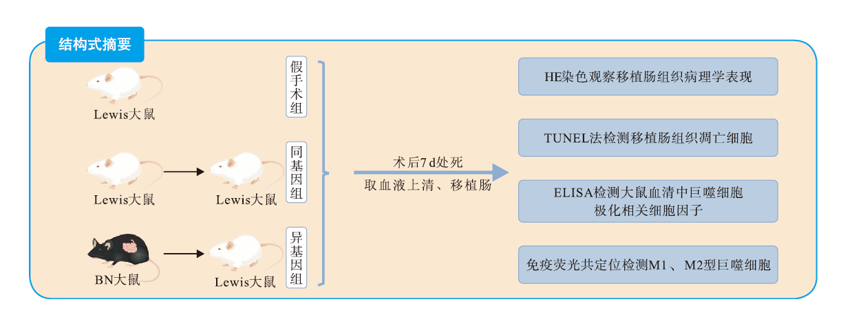
目的 探究小肠移植术后发生急性排斥反应(AR)时巨噬细胞极化状态的改变。 方法 将6只Brown Norway(BN)大鼠和24只Lewis大鼠分为假手术组(6只Lewis大鼠)、同基因组(Lewis→Lewis,供受体各6只)和异基因组(BN→Lewis,供受体各6只)。对各组大鼠术后7 d的移植肠组织进行苏木素-伊红(HE)染色和脱氧核糖核酸末端转移酶介导的 dUTP 缺口末端标记(TUNEL)法检测,观察其病理学表现和细胞凋亡情况;采用酶联免疫吸附试验(ELISA)检测血清中M1和M2型巨噬细胞极化相关细胞因子表达水平;利用免疫荧光技术检测各组移植肠组织中M1和M2型巨噬细胞表面标志物并进行共定位计数分析。 结果 HE染色和TUNEL检测结果显示假手术组与同基因组肠上皮形态结构正常,未见明显凋亡小体;异基因组大鼠术后7 d移植肠组织上皮层绒毛结构破坏严重,隐窝数量减少,凋亡小体增多,炎症细胞浸润肠壁全层,呈现中-重度AR。ELISA结果显示异基因组受体鼠血清中M1型巨噬细胞极化相关细胞因子肿瘤坏死因子(TNF)-α、干扰素(IFN)-γ和白细胞介素(IL)-12表达水平高于假手术组和同基因组,同基因组中M2型巨噬细胞极化相关细胞因子IL-10和转化生长因子(TGF)-β表达水平高于假手术组和异基因组,差异均有统计学意义(均为P<0.05)。免疫荧光结果显示异基因组移植肠组织中M1型巨噬细胞计数多于假手术组和同基因组,同基因组M2型巨噬细胞计数多于假手术组和异基因组,差异均有统计学意义(均为P<0.05)。 结论 小肠移植术后发生AR的移植物中,大量巨噬细胞浸润肠壁全层,以M1型为主并分泌大量促炎因子,调控巨噬细胞极化方向是治疗小肠移植术后AR的潜在方法。 Abstract:Objective To investigate the changes of macrophage polarization during acute rejection (AR) after intestinal transplantation. Methods Six Brown Norway (BN) rats and 24 Lewis rats were divided into the sham operation group (6 Lewis rats), syngeneic transplantation group (Lewis→Lewis, 6 donors and 6 recipients) and allogeneic transplantation group (BN→Lewis, 6 donors and 6 recipients). At postoperative 7 d, the intestinal graft tissues in all groups were collected for hematoxylin-eosin (HE) staining and terminal deoxynucleotidyl transferase-mediated dUTP nick-end labeling (TUNEL) assay. Pathological manifestations and cell apoptosis were observed. The expression levels of serum cytokines related to M1 and M2 macrophage polarization were determined by enzyme-linked immunosorbent assay (ELISA). Surface markers of M1 and M2 macrophages of intestinal graft tissues in each group were co-localized and counted by immunofluorescence staining. Results HE staining and TUNEL assay showed that the intestinal epithelial morphology and structure were normal and no evident apoptotic bodies were found in the sham operation and syngeneic transplantation groups. At 7 d after transplantation, the epithelial villi structure of intestinal graft tissues was severely damaged, the number of crypts was decreased, the number of apoptotic bodies was increased, and inflammatory cells infiltrated into the whole intestinal wall, manifested with moderate to severe AR in the allogeneic transplantation group. ELISA revealed that the expression levels of serum cytokines related to M1 macrophage polarization, such as tumor necrosis factor (TNF)-α, interferon (IFN)-γ and interleukin (IL)-12, of the recipient rats in the allogeneic transplantation group were higher than those in the sham operation and syngeneic transplantation groups. The expression levels of serum cytokines related to M2 macrophage polarization, such as IL-10 and transforming growth factor (TGF)-β, in the syngeneic transplantation group were higher compared with those in the sham operation and allogeneic transplantation group, and the differences were statistically significant (all P<0.05). Immunofluorescence staining showed that the number of M1 macrophages in the allogeneic transplantation group was higher than those in the sham operation and syngeneic transplantation groups, and the number of M2 macrophages in the syngeneic transplantation group was higher than those in the sham operation and allogeneic transplantation groups, and the differences were statistically significant (all P<0.05). Conclusions Among the allografts with AR after intestinal transplantation, a large number of macrophages, mainly M1 macrophages secreting a large number of pro-inflammatory cytokines, infiltrate into the whole intestinal wall. Regulating the direction of macrophage polarization is a potential treatment for AR after intestinal transplantation. -
表 1 各组血清中M1型与M2型巨噬细胞极化相关细胞因子表达水平($\bar {\boldsymbol{x}} $±s,pg/mL)
Table 1. Expression levels of M1 and M2 macrophage polarization related cytokines in serum of each group
组别 n TNF-α IFN-γ IL-12 IL-10 TGF-β 假手术组 6 89.3±2.5 47.4±5.0 10.3±0.6 23. 8±1.0 97.7±4.8 同基因组 6 97.9±6.1 50.0±2.1 11.1±1.2 45.8±1.9a 147.1±19.2a 异基因组 6 213.8±4.1a,b 102.4±3.3 a,b 22.1±1.3 a,b 36.6±2.1b 74.6±3.6b F值 1 456.16 432.27 234.97 246.95 60.99 注:与假手术组比较,aP<0.05,与同基因组比较,bP<0.05。 -
[1] 吴国生,梁廷波. 自体小肠移植技术的实践与挑战[J]. 中华消化外科杂志, 2021, 20(1): 85-88. DOI: 10.3760/cma.j.cn115610-20201202-00748.WU GS, LIANG TB. Current practice and challenges of intestinal autotransplantation [J]. Chin J Dig Surg, 2021, 20(1): 85-88. DOI: 10.3760/cma.j.cn115610-20201202-00748. [2] PETERSON LW, ARTIS D. Intestinal epithelial cells: regulators of barrier function and immune homeostasis[J]. Nat Rev Immunol, 2014, 14(3): 141-153. DOI: 10.1038/nri3608. [3] MAO K, BAPTISTA AP, TAMOUTOUNOUR S, et al. Innate and adaptive lymphocytes sequentially shape the gut microbiota and lipid metabolism[J]. Nature, 2018, 554(7691): 255-259. DOI: 10.1038/nature25437. [4] ROBERTS MB, FISHMAN JA. Immunosuppressive agents and infectious risk in transplantation: managing the "net state of immunosuppression"[J]. Clin Infect Dis, 2021, 73(7): e1302-e1317. DOI: 10.1093/cid/ciaa1189. [5] MIYAGAWA S, KODAMA T, MATSUURA R, et al. A study of the mechanisms responsible for the action of new immunosuppressants and their effects on rat small intestinal transplantation[J]. Transpl Immunol, 2022, 70: 101497. DOI: 10.1016/j.trim.2021.101497. [6] DOGRA H, HIND J. Innovations in immunosuppression for intestinal transplantation[J]. Front Nutr, 2022, 9: 869399. DOI: 10.3389/fnut.2022.869399. [7] FLANNIGAN KL, GEEM D, HARUSATO A, et al. Intestinal antigen-presenting cells: key regulators of immune homeostasis and inflammation[J]. Am J Pathol, 2015, 185(7): 1809-1819. DOI: 10.1016/j.ajpath.2015.02.024. [8] MOREIRA LOPES TC, MOSSER DM, GONÇALVES R. Macrophage polarization in intestinal inflammation and gut homeostasis[J]. Inflamm Res, 2020, 69(12): 1163-1172. DOI: 10.1007/s00011-020-01398-y. [9] LI C, XU MM, WANG K, et al. Macrophage polarization and meta-inflammation[J]. Transl Res, 2018, 191: 29-44. DOI: 10.1016/j.trsl.2017.10.004. [10] VIOLA MF, BOECKXSTAENS G. Niche-specific functional heterogeneity of intestinal resident macrophages[J]. Gut, 2021, 70(7): 1383-1395. DOI: 10.1136/gutjnl-2020-323121. [11] YE L, HE S, MAO X, et al. Effect of hepatic macrophage polarization and apoptosis on liver ischemia and reperfusion injury during liver transplantation[J]. Front Immunol, 2020, 11: 1193. DOI: 10.3389/fimmu.2020.01193. [12] XU XS, FENG ZH, CAO D, et al. SCARF1 promotes M2 polarization of Kupffer cells via calcium-dependent PI3K-Akt-STAT3 signalling to improve liver transplantation[J]. Cell Prolif, 2021, 54(4): e13022. DOI: 10.1111/cpr.13022. [13] MÖLNE J, NASIC S, BRÖCKER V, et al. Glomerular macrophage index (GMI) in kidney transplant biopsies is associated with graft outcome[J]. Clin Transplant, 2022, 36(12): e14816. DOI: 10.1111/ctr.14816. [14] 任滌非, 廖涛, 苗芸. 巨噬细胞在移植后慢性排斥反应中的作用研究进展[J]. 器官移植, 2023, 14(3): 358-363. DOI: 10.3969/j.issn.1674-7445.2023.03.006.REN DF, LIAO T, MIAO Y. Research progress on the role of macrophages in post-transplantation chronic rejection[J]. Organ Transplant, 2023, 14(3): 358-363. DOI: 10.3969/j.issn.1674-7445.2023.03.006. [15] 董博清, 李杨, 石玉婷, 等. 基于加权基因共表达网络鉴定肾移植术后排斥反应中巨噬细胞M1亚型相关基因[J]. 器官移植, 2023, 14(1): 83-92. DOI: 10.3969/j.issn.1674-7445.2023.01.011.DONG BQ, LI Y, SHI YT, et al. Identification of M1 macrophage-related genes in rejection after kidney transplantation based on weighted gene co-expression network analysis[J]. Organ Transplant, 2023, 14(1): 83-92. DOI: 10.3969/j.issn.1674-7445.2023.01.011. [16] KOPECKY BJ, FRYE C, TERADA Y, et al. Role of donor macrophages after heart and lung transplantation[J]. Am J Transplant, 2020, 20(5): 1225-1235. DOI: 10.1111/ajt.15751. [17] GAO C, WANG X, LU J, et al. Mesenchymal stem cells transfected with sFGL2 inhibit the acute rejection of heart transplantation in mice by regulating macrophage activation[J]. Stem Cell Res Ther, 2020, 11(1): 241. DOI: 10.1186/s13287-020-01752-1. [18] KOPECKY BJ, DUN H, AMRUTE JM, et al. Donor macrophages modulate rejection after heart transplantation[J]. Circulation, 2022, 146(8): 623-638. DOI: 10.1161/CIRCULATIONAHA.121.057400. [19] TIAN H, WU J, MA M. Implications of macrophage polarization in corneal transplantation rejection[J]. Transpl Immunol, 2021, 64: 101353. DOI: 10.1016/j.trim.2020.101353. [20] TOYAMA C, MAEDA A, KOGATA S, et al. Effect of a C5a receptor antagonist on macrophage function in an intestinal transplant rat model[J]. Transpl Immunol, 2022, 72: 101559. DOI: 10.1016/j.trim.2022.101559. [21] FOELL D, BECKER F, HADRIAN R, et al. A practical guide for small bowel transplantation in rats-review of techniques and models[J]. J Surg Res, 2017, 213: 115-130. DOI: 10.1016/j.jss.2017.02.026. [22] 郭晖, 陈知水. 移植小肠病理学诊断标准及其进展[J]. 器官移植, 2022, 13(3): 307-316. DOI: 10.3969/j.issn.1674-7445.2022.03.005.GUO H, CHEN ZS. Diagnostic criteria and its progress on intestinal graft pathology [J]. Organ Transplant, 2022, 13(3): 307-316. DOI: 10.3969/j.issn.1674-7445.2022.03.005. [23] GHARRAEE N, WANG Z, PFLUM A, et al. Eicosapentaenoic acid ameliorates cardiac fibrosis and tissue inflammation in spontaneously hypertensive rats[J]. J Lipid Res, 2022, 63(11): 100292. DOI: 10.1016/j.jlr.2022.100292. [24] OGINO T, TAKEDA K. Immunoregulation by antigen-presenting cells in human intestinal lamina propria[J]. Front Immunol, 2023, 14: 1138971. DOI: 10.3389/fimmu.2023.1138971. [25] MOKARRAM N, DYMANUS K, SRINIVASAN A, et al. Immunoengineering nerve repair[J]. Proc Natl Acad Sci U S A, 2017, 114(26): E5077-E5084. DOI: 10.1073/pnas.1705757114. [26] MINUTTI CM, JACKSON-JONES LH, GARCÍA-FOJEDA B, et al. Local amplifiers of IL-4Rα-mediated macrophage activation promote repair in lung and liver[J]. Science, 2017, 356(6342): 1076-1080. DOI: 10.1126/science.aaj2067. [27] HUANG H, ZHANG X, ZHANG C, et al. The time-dependent shift in the hepatic graft and recipient macrophage pool following liver transplantation[J]. Cell Mol Immunol, 2020, 17(4): 412-414. DOI: 10.1038/s41423-019-0253-x. [28] CAO ZR, ZHENG WX, JIANG YX, et al. miR-449a ameliorates acute rejection after liver transplantation via targeting procollagen-lysine1, 2-oxoglutarate5-dioxygenase 1 in macrophages[J]. Am J Transplant, 2023, 23(3): 336-352. DOI: 10.1016/j.ajt.2022.12.009. [29] AZAD TD, DONATO M, HEYLEN L, et al. Inflammatory macrophage-associated 3-gene signature predicts subclinical allograft injury and graft survival[J]. JCI Insight, 2018, 3(2): e95659. DOI: 10.1172/jci.insight.95659. [30] HANG Z, WEI J, ZHENG M, et al. Iguratimod attenuates macrophage polarization and antibody-mediated rejection after renal transplant by regulating KLF4[J]. Front Pharmacol, 2022, 13: 865363. DOI: 10.3389/fphar.2022.865363. [31] ZHAO HY, LYU ZS, DUAN CW, et al. An unbalanced monocyte macrophage polarization in the bone marrow microenvironment of patients with poor graft function after allogeneic haematopoietic stem cell transplantation[J]. Br J Haematol, 2018, 182(5): 679-692. DOI: 10.1111/bjh.15452. [32] 张翔, 王子杰, 郑明, 等. M1型巨噬细胞极化在内皮细胞转分化及慢性移植肾失功中的作用[J]. 南京医科大学学报(自然科学版), 2021, 41(9): 1296-1303, 1309. DOI: 10.7655/NYDXBNS20210904.ZHAGN X, WANG ZJ, ZHENG M, et al. The role of M1 polarized-macrophage in endothelial-to-myofibroblast transition and chronic allograft dysfunction[J]. J Nanjing Med Univ(Nat sci), 2021, 41(9): 1296-1303, 1309. DOI: 10.7655/NYDXBNS20210904. [33] 简迅, 王丹阳, 许燕楠, 等. 极化骨髓巨噬细胞移植对CCl4诱导的肝纤维化大鼠模型的影响[J]. 临床肝胆病杂志, 2021, 37(12): 2830-2837. DOI: 10.3969/j.issn.1001-5256.2021.12.020.JIAN X, WANG DY, XU YN, et al. Effect of polarized bone marrow -derived macrophage transplantation on the progression of CCl4 -induced liver fibrosis in rats[J]. J Clin Hepatol, 2021, 37(12): 2830-2837. DOI: 10.3969/j.issn.1001-5256.2021.12.020. [34] YU J, LI P, LI Z, et al. Topical administration of 0.3% tofacitinib suppresses M1 macrophage polarization and allograft corneal rejection by blocking STAT1 activation in the rat cornea[J]. Transl Vis Sci Technol, 2022, 11(3): 34. DOI: 10.1167/tvst.11.3.34. [35] HANAKI R, TOYODA H, IWAMOTO S, et al. Donor-derived M2 macrophages attenuate GVHD after allogeneic hematopoietic stem cell transplantation[J]. Immun Inflamm Dis, 2021, 9(4): 1489-1499. DOI: 10.1002/iid3.503. -


 下载:
下载:


