Effect of liver transplantation on intestinal microflora in children with biliary atresia
-
摘要:
目的 研究肝移植对胆道闭锁儿童肠道菌群的影响。 方法 收集16例胆道闭锁儿童肝移植术前、术后6个月的粪便样本及肝功能指标等临床数据,10名健康儿童作为健康对照组。对粪便样本进行DNA提取和宏基因组测序,采用R语言等软件包进行数据分析。分析肝移植术后肠道菌群物种结构及功能组成的改变情况,检测胆道闭锁儿童肝移植术后肠道菌群恢复的程度,分析肠道菌群与肝功能指标间的关系。 结果 肝移植术后,胆道闭锁儿童肠道菌群的物种数量增加。胆道闭锁儿童肝移植术前肠道菌群以机会致病菌为主,移植术后机会致病菌丰度下降,产短链脂肪酸菌丰度升高(均为P < 0.05)。肝移植术后,脂代谢、氨基酸代谢、碳水化合物代谢、能量代谢、辅助因子和维生素代谢等代谢通路增强,而细菌感染、免疫系统疾病、药物抵抗等相关代谢通路减弱。与健康对照组比较,肝移植术后组肠道菌群多样性和菌群结构差异均无统计学意义,但两组间仍存在差异物种。胆道闭锁儿童肝移植术后肝功能指标均呈下降趋势(均为P < 0.000 1)。肠道有益菌的丰度与肝功能指标呈负相关,而机会致病菌丰度与肝功能指标呈正相关(均为P < 0.05)。 结论 肝移植在一定程度上可以显著改善胆道闭锁儿童肠道菌群的结构及功能组成。 Abstract:Objective To evaluate the effect of liver transplantation on intestinal microflora in children with biliary atresia. Methods The fecal samples and liver function indexes of 16 children with biliary atresia before and 6 months after liver transplantation were collected, and 10 healthy children were selected as the healthy controls. DNA extraction and metagenome sequencing were carried out in the fecal samples. Statistical analysis was performed by software packages, such as R language. The changes of species structure and functional composition of intestinal microflora after liver transplantation were analyzed. The recovery of intestinal microflora in children with biliary atresia after liver transplantation was assessed. The relationship between intestinal microflora and liver function indexes was investigated. Results Following liver transplantation, the number of species of intestinal microflora in children with biliary atresia was increased. The opportunistic pathogens were the dominant species of intestinal microflora in children with biliary atresia before liver transplantation. The abundance of opportunistic pathogens was decreased and the abundance of short-chain fatty acid-producing bacteria was increased after liver transplantation (all P < 0.05). Following liver transplantation, lipid metabolism, amino acid metabolism, carbohydrate metabolism, energy metabolism, metabolism of cofactors and vitamins were enhanced, whereas infectious diseases of bacterial, immune diseases and drug resistance were weakened. Compared with the healthy control group, there were no statistically significant differences in the diversity and structure of intestinal microflora in the post-liver transplant group, but different species were observed between two groups. The liver function indexes of children with biliary atresia after liver transplantation tended to decline (all P < 0.000 1). The abundance of beneficial intestinal microflora was negatively correlated with liver function indexes, whereas the abundance of opportunistic pathogens was positively correlated with liver function indexes (all P < 0.05). Conclusions Liver transplantation may significantly improve the structure and functional composition of intestinal microflora in children with biliary atresia. -

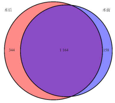
图 1 肝移植术前组和肝移植术后组肠道菌群物种数量的韦恩图(属水平)
Figure 1. Venn diagram of the number of intestinal microflora species in the pre- and post-liver transplant group(genus level)

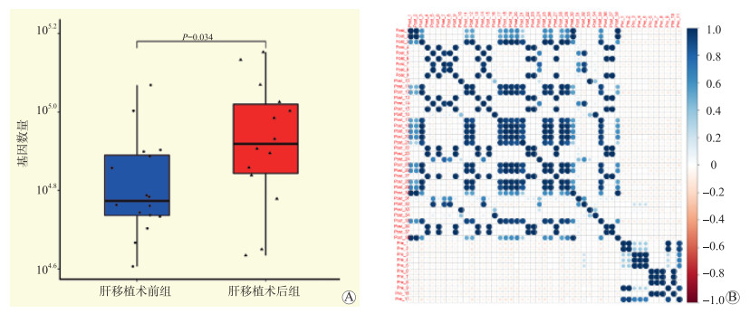
图 2 肝移植术前组和肝移植术后组肠道菌群基因数量比较及相关性分析
注:A图为肝移植术前组和肝移植术后组基因数量比较;B图为两组基因间的Spearman相关性分析(仅显示P < 0.05的结果)。
Figure 2. Comparison and correlation analysis of the number of intestinal microflora genes in the pre- and post-liver transplant group

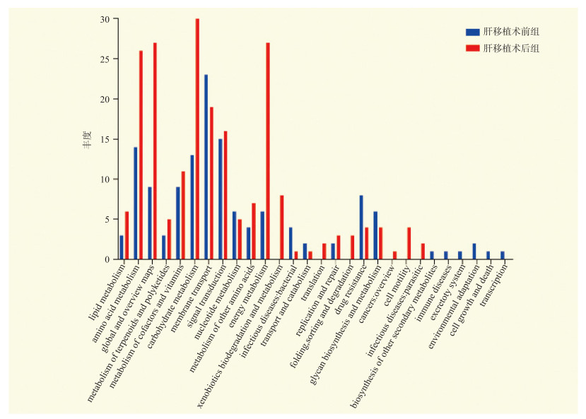
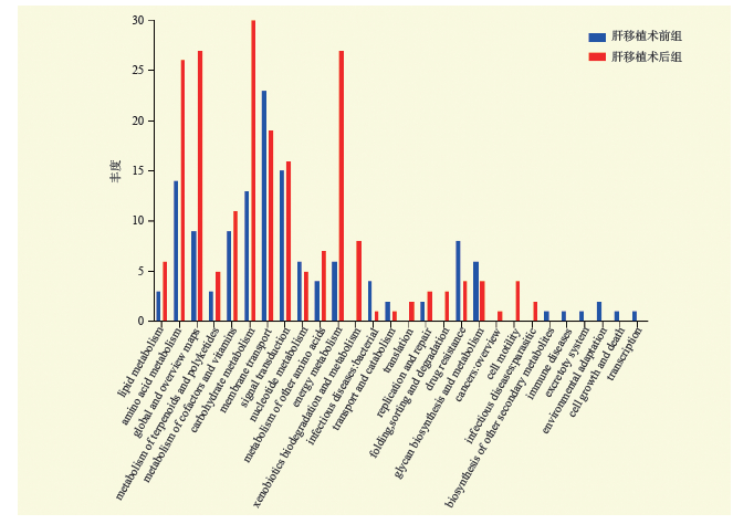
图 3 肝移植术前组和肝移植术后组肠道菌群功能代谢通路比较
Figure 3. Comparison of functional metabolic pathways of intestinal microflora between the pre- and post-liver transplant group

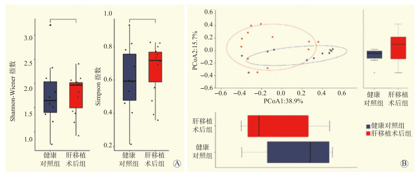
图 4 肝移植术后组与健康对照组肠道菌群多样性和菌群结构比较
注:A图为肝移植术后组与健康对照组肠道菌群α多样性比较;B图为肝移植术后组与健康对照组肠道菌群β多样性比较,横纵坐标的刻度为相对距离,无实际意义,PCoA1表示尽可能最大解释数据变化的主坐标成分,PCoA1(a%)表示第一主坐标所占比例为a%,PCoA2表示剩余的变化度中占比例最大的主坐标成分。
Figure 4. Comparison of intestinal microflora diversity and structure between post-liver transplant group and healthy control group

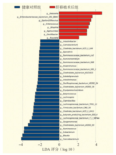
图 5 肝移植术后组与健康对照组差异物种比较
Figure 5. Comparison of species between post-liver transplant group and healthy control group

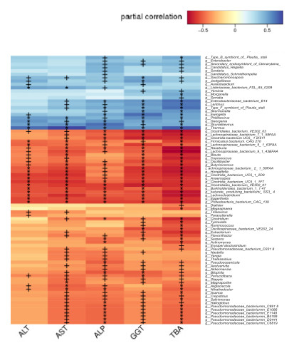
图 6 肠道菌群与肝功能指标的相关性分析热图
注:图中颜色的深浅直观展示物种与肝功能指标的相关性大小,蓝色代表正相关,红色代表负相关,*为P < 0.05,+为P < 0.01。
Figure 6. Heat map of correlation analysis between intestinal microflora and liver function indexes
表 1 肝移植术前组和肝移植术后组丰度发生改变的物种(属水平)
Table 1. Species with altered abundance in the pre- and post-liver transplant group(genus level)
物种名称 肝移植术后组(n=16) 肝移植术前组(n=16) P值 富集组 g__Enterobacter 0.000 940 0 0.007 170 0 0.040 术前 g__Yersinia 0.000 039 0 0.000 325 0 0.030 术前 g__Morganella 0.000 017 0 0.000 090 0 0.010 术前 g__Serratia 0.000 017 0 0.000 038 0 0.040 术前 g__Blautia 0.004 100 0 0.000 058 0 0.003 术后 g__Clostridium 0.003 050 0 0.000 400 0 0.007 术后 g__Eubacterium 0.001 200 0 0.000 030 0 0.002 术后 g__Roseburia 0.000 380 0 0.000 010 0 0.001 术后 g__Akkermansia 0.000 007 0 0.000 000 9 0.030 术后  下载: 导出CSV
表 2 肝移植术前组和肝移植术后组肝功能指标比较
Table 2. Comparison of liver function indexes between pre- and post-liver transplant group[M(Q25, Q75)]
肝功能指标 肝移植术前组(n=16) 肝移植术后组(n=16) P值 ALT(U/L) 151(62,181) 21(17,33) < 0.000 1 AST(U/L) 212(107,272) 36(27,40) < 0.000 1 ALP(U/L) 611(434,833) 249(204,329) < 0.000 1 GGT(U/L) 181(106,532) 17(14,33) < 0.000 1 TBA(μmol/L) 164(88,284) 9(5,13) < 0.000 1 TB(μmol/L) 281(8,557) 8(5,10) < 0.000 1
下载: 导出CSV
-
[1] 殷润开, 赵瑞芹, 付海燕, 等. 肝功能相关指标诊断婴儿胆道闭锁的价值[J]. 实用医学杂志, 2021, 37(12): 1607-1612. DOI: 10.3969/j.issn.1006-5725.2021.12.019.YIN RK, ZHAO RQ, FU HY, et al. Value of liver function index in the diagnosis of biliary atresia in infants[J]. J Pract Med, 2021, 37(12): 1607-1612. DOI: 10.3969/j.issn.1006-5725.2021.12.019. [2] SOKOL RJ, SHEPHERD RW, SUPERINA R, et al. Screening and outcomes in biliary atresia: summary of a National Institutes of Health workshop[J]. Hepatology, 2007, 46(2): 566-581. DOI: 10.1002/hep.21790. [3] SIDDIQUI AI, AHMAD T. Biliary atresia[M]. Treasure Island (FL): StatPearls, 2021. [4] KASAHARA M, UMESHITA K, SAKAMOTO S, et al. Liver transplantation for biliary atresia: a systematic review[J]. Pediatr Surg Int, 2017, 33(12): 1289-1295. DOI: 10.1007/s00383-017-4173-5. [5] WEI Y, LI Y, YAN L, et al. Alterations of gut microbiome in autoimmune hepatitis[J]. Gut, 2020, 69(3): 569-577. DOI: 10.1136/gutjnl-2018-317836. [6] HU H, LIN A, KONG M, et al. Intestinal microbiome and NAFLD: molecular insights and therapeutic perspectives[J]. J Gastroenterol, 2020, 55(2): 142-158. DOI: 10.1007/s00535-019-01649-8. [7] CASSARD AM, CIOCAN D. Microbiota, a key player in alcoholic liver disease[J]. Clin Mol Hepatol, 2018, 24(2): 100-107. DOI: 10.3350/cmh.2017.0067. [8] MILOSEVIC I, VUJOVIC A, BARAC A, et al. Gut-liver axis, gut microbiota, and its modulation in the management of liver diseases: a review of the literature[J]. Int J Mol Sci, 2019, 20(2): 395. DOI: 10.3390/ijms20020395. [9] MOHAJERI MH, BRUMMER RJM, RASTALL RA, et al. The role of the microbiome for human health: from basic science to clinical applications[J]. Eur J Nutr, 2018, 57(Suppl 1): 1-14. DOI: 10.1007/s00394-018-1703-4. [10] XING HC, LI LJ, XU KJ, et al. Protective role of supplement with foreign bifidobacterium and lactobacillus in experimental hepatic ischemia-reperfusion injury[J]. J Gastroenterol Hepatol, 2006, 21(4): 647-656. DOI: 10.1111/j.1440-1746.2006.04306.x. [11] ZHU B, WANG X, LI L. Human gut microbiome: the second genome of human body[J]. Protein Cell, 2010, 1(8): 718-725. DOI: 10.1007/s13238-010-0093-z. [12] WANG J, QIAN T, JIANG J, et al. Gut microbial profile in biliary atresia: a case-control study[J]. J Gastroenterol Hepatol, 2020, 35(2): 334-342. DOI: 10.1111/jgh.14777. [13] ASAI A, MIETHKE A, BEZERRA JA. Pathogenesis of biliary atresia: defining biology to understand clinical phenotypes[J]. Nat Rev Gastroenterol Hepatol, 2015, 12(6): 342-352. DOI: 10.1038/nrgastro.2015.74. [14] ZENG Y, CHEN S, FU Y, et al. Gut microbiota dysbiosis in patients with hepatitis B virus-induced chronic liver disease covering chronic hepatitis, liver cirrhosis and hepatocellular carcinoma[J]. J Viral Hepat, 2020, 27(2): 143-155. DOI: 10.1111/jvh.13216. [15] LV LX, FANG DQ, SHI D, et al. Alterations and correlations of the gut microbiome, metabolism and immunity in patients with primary biliary cirrhosis[J]. Environ Microbiol, 2016, 18(7): 2272-2286. DOI: 10.1111/1462-2920.13401. [16] WENG MT, CHIU YT, WEI PY, et al. Microbiota and gastrointestinal cancer[J]. J Formos Med Assoc, 2019, 118(Suppl 1): S32-S41. DOI: 10.1016/j.jfma.2019.01.002. [17] TANG R, WEI Y, LI Y, et al. Gut microbial profile is altered in primary biliary cholangitis and partially restored after UDCA therapy[J]. Gut, 2018, 67(3): 534-541. DOI: 10.1136/gutjnl-2016-313332. [18] ABE K, FUJITA M, HAYASHI M, et al. Gut and oral microbiota in autoimmune liver disease[J]. Fukushima J Med Sci, 2020, 65(3): 71-75. DOI: 10.5387/fms.2019-21. [19] TRIPATHI A, DEBELIUS J, BRENNER DA, et al. The gut-liver axis and the intersection with the microbiome[J]. Nat Rev Gastroenterol Hepatol, 2018, 15(7): 397-411. DOI: 10.1038/s41575-018-0011-z. [20] ISAACS-TEN A, ECHEANDIA M, MORENO-GONZALEZ M, et al. Intestinal microbiome-macrophage crosstalk contributes to cholestatic liver disease by promoting intestinal permeability in mice[J]. Hepatology, 2020, 72(6): 2090-2108. DOI: 10.1002/hep.31228. [21] YANG X, LU D, ZHUO J, et al. The gut-liver axis in immune remodeling: new insight into liver diseases[J]. Int J Biol Sci, 2020, 16(13): 2357-2366. DOI: 10.7150/ijbs.46405. [22] ISLAM KB, FUKIYA S, HAGIO M, et al. Bile acid is a host factor that regulates the composition of the cecal microbiota in rats[J]. Gastroenterology, 2011, 141(5): 1773-1781. DOI: 10.1053/j.gastro.2011.07.046. [23] LONG SL, GAHAN CGM, JOYCE SA. Interactions between gut bacteria and bile in health and disease[J]. Mol Aspects Med, 2017, 56: 54-65. DOI: 10.1016/j.mam.2017.06.002. [24] BAJAJ JS, FAGAN A, SIKAROODI M, et al. Liver transplant modulates gut microbial dysbiosis and cognitive function in cirrhosis[J]. Liver Transpl, 2017, 23(7): 907-914. DOI: 10.1002/lt.24754. [25] BAJAJ JS, KAKIYAMA G, COX IJ, et al. Alterations in gut microbial function following liver transplant[J]. Liver Transpl, 2018, 24(6): 752-761. DOI: 10.1002/lt.25046. [26] NORVELL JP. Spotlight on impactful research: impact of liver transplantation on gut microbiota and cognitive function[J]. Clin Liver Dis (Hoboken), 2019, 13(3): 72-73. DOI: 10.1002/cld.746. [27] PILLAI AA, LEVITSKY J. Overview of immunosuppression in liver transplantation[J]. World J Gastroenterol, 2009, 15(34): 4225-4233. DOI: 10.3748/wjg.15.4225. [28] SUN LY, YANG YS, QU W, et al. Gut microbiota of liver transplantation recipients[J]. Sci Rep, 2017, 7(1): 3762. DOI: 10.1038/s41598-017-03476-4. [29] ZHANG J, REN FG, LIU P, et al. Characteristics of fecal microbial communities in patients with non-anastomotic biliary strictures after liver transplantation[J]. World J Gastroenterol, 2017, 23(46): 8217-8226. DOI: 10.3748/wjg.v23.i46.8217. [30] HOLOTA Y, DOVBYNCHUK T, KAJI I, et al. The long-term consequences of antibiotic therapy: role of colonic short-chain fatty acids (SCFA) system and intestinal barrier integrity[J]. PLoS One, 2019, 14(8): e0220642. DOI: 10.1371/journal.pone.0220642. [31] AGUS A, CLÉMENT K, SOKOL H. Gut microbiota-derived metabolites as central regulators in metabolic disorders[J]. Gut, 2021, 70(6): 1174-1182. DOI: 10.1136/gutjnl-2020-323071. -

 下载:
下载:

 点击查看大图
计量
- 文章访问数: 222
- HTML全文浏览量: 60
- PDF下载量: 72
- 被引次数: 0