Effect of induced pluripotent stem cell transplantation on treatment of cerebral ischemic injury in rats after down-regulation of HIF-2α gene
-
摘要:
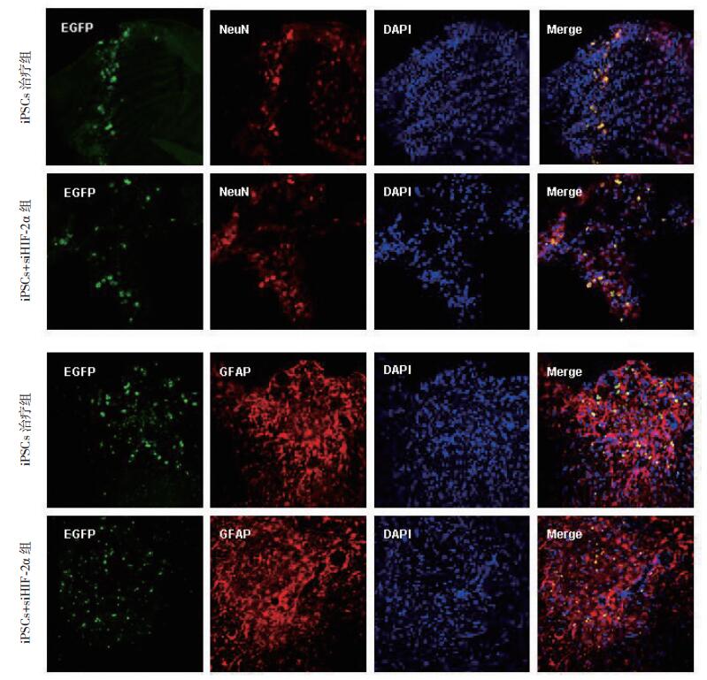
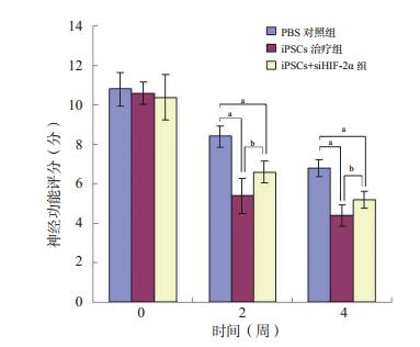
目的 探讨低氧诱导因子(HIF)-2α基因下调后的诱导多能干细胞(iPSCs)移植对大鼠脑缺血损伤治疗作用的影响。 方法 24只雄性SD大鼠随机分为3组(每组8只):iPSCs治疗组、iPSCs+siHIF-2α组,磷酸盐缓冲液(PBS)对照组。所有大鼠均采用大脑中动脉栓塞法制作脑缺血模型,栓塞90 min后进行再灌注。再灌注后3 d进行iPSCs移植、iPSCs+siHIF-2α移植或PBS注射。在移植之前及之后的第2、4周,行神经行为学评价并采用小动物正电子发射体层仪(micro-PET)扫描测定18F-氟代脱氧葡萄糖(18F-FDG)的摄取值。移植后第4周,处死大鼠行免疫荧光检测神经细胞标志物。 结果 在干细胞移植后第2、4周,iPSCs治疗组的神经功能评分均明显低于PBS对照组(均为P < 0.05),而iPSCs+siHIF-2α组的神经功能评分在这两个时间点均高于iPSCs治疗组(均为P < 0.05)。在干细胞移植后的第2、4周,与PBS对照组比较,iPSCs治疗组和iPSC+siHIF-2α组脑缺血大鼠脑部的糖代谢水平(损伤侧/正常侧)明显增加(均为P < 0.001)。干细胞移植后第2周,iPSCs治疗组大鼠脑缺血区糖代谢水平明显高于iPSCs+siHIF-2α组(P < 0.001)。此后iPSCs+siHIF-2α组大鼠脑缺血区糖代谢水平持续上升,至干细胞移植后第4周,与iPSCs治疗组较为接近,但仍低于iPSCs治疗组(P=0.025)。免疫荧光结果提示移植的干细胞存活并迁移至梗死区周边,并且大部分的移植干细胞均表达了神经细胞标志物。 结论 iPSCs移植对大鼠脑缺血损伤有明显的治疗作用,HIF-2α基因下调对iPSCs的治疗作用有一定的影响。 -
关键词:
- 脑缺血 /
- 诱导多功能干细胞 /
- 干细胞移植 /
- 低氧诱导因子-2α /
- 小动物正电子发射体层仪(micro-PET) /
- 18F-氟代脱氧葡萄糖(18F-FDG) /
- 糖代谢 /
- 小干扰核糖核酸(siRNA)
Abstract:Objective To investigate the effect of induced pluripotent stem cells (iPSCs) transplantation after the down-regulation of hypoxia-inducible factor (HIF)-2α gene on the treatment of cerebral ischemia injury in rats. Methods Twenty-four male Sprague-Dawley (SD) rats were randomly and evenly divided into the iPSCs treatment, iPSCs+siHIF-2α and phosphate buffer saline (PBS) control groups. The rat models with cerebral ischemia were established by the middle cerebral artery embolization, and reperfusion was carried out 90 min after embolization. At 3 d after reperfusion, iPSCs transplantation, iPSCs+siHIF-2α transplantation or PBS injection were performed. Before and at 2 and 4 weeks after transplantation, neurobehavioral evaluation was performed and the uptake of 18F-fluoro-deoxyglucose (18F-FDG) was measured by micro-positron emission tomography (micro-PET). At 4 weeks after transplantation, the rats were sacrificed and nerve cell markers were detected by immunofluorescence. Results At 2 and 4 weeks after stem cell transplantation, the neurological function scores of the iPSCs treatment group were significantly lower than those in the PBS control group (both P < 0.05), whereas the neurological function scores in the iPSCs+siHIF-2α group were considerably higher than those in the iPSCs treatment group (both P < 0.05). At 2 and 4 weeks after stem cell transplantation, the glucose metabolism levels (injured side/normal side) in the brain of cerebral ischemia rats in the iPSCs treatment and iPSCs+siHIF-2αgroups were significantly increased compared with those in the PBS control group (both P < 0.001). At the 2nd week after stem cell transplantation, the glucose metabolism level in the iPSCs treatment group was significantly higher than that in the iPSCs+siHIF-2α group (P < 0.001). Subsequently, the glucose metabolism level in the iPSCs+siHIF-2α group continued to rise until the 4th week after stem cell transplantation, which was almost identical to but still lower than that in the iPSCs treatment group (P=0.025). Immunofluorescent staining prompted that the transplanted stem cells survived and migrated to the periphery of the infarction area, and a majority of the transplanted stem cells expressed nerve cell markers. Conclusions iPSCs transplantation can be employed to effectively treat cerebral ischemia injury in rats. Down-regulation of HIF-2α gene exerts certain effect upon the therapeutic effect of iPSCs transplantation. -

图 1 各组大鼠干细胞移植前后神经功能评分的比较
与PBS对照组比较,aP < 0.05;与iPSCs治疗组比较,bP < 0.05
Figure 1. Comparison of neural function scores in rats among each group before and after stem cell transplantation

图 2 各组大鼠干细胞移植前后脑缺血区糖代谢的变化
Figure 2. Change of glucose metabolism in cerebral ischemia area of rats in each group before and after stem cell transplantation
-
[1] FUJIMOTO Y, ABEMATSU M, FALK A, et al. Treatment of a mouse model of spinal cord injury by transplantation of human induced pluripotent stem cell-derived long-term self-renewing neuroepithelial-like stem cells[J]. Stem Cells, 2012, 30(6): 1163-1173. DOI: 10.1002/stem.1083. [2] GLASS JD, BOULIS NM, JOHE K, et al. Lumbar intraspinal injection of neural stem cells in patients with amyotrophic lateral sclerosis: results of a phase Ⅰ trial in 12 patients[J]. Stem Cells, 2012, 30(6): 1144-1151. DOI: 10.1002/stem.1079. [3] CUSIMANO M, BIZIATO D, BRAMBILLA E, et al. Transplanted neural stem/precursor cells instruct phagocytes and reduce secondary tissue damage in the injured spinal cord[J]. Brain, 2012, 135(Pt 2): 447-460. DOI: 10.1093/brain/awr339. [4] AL-ZOUBI A, JAFAR E, JAMOUS M, et al. Transplantation of purified autologous leukapheresis-derived CD34+ and CD133+ stem cells for patients with chronic spinal cord injuries: long-term evaluation of safety and efficacy[J]. Cell Transplant, 2014, 23(Suppl 1): S25-S34. DOI: 10.3727/096368914X684899. [5] FELDMAN EL, BOULIS NM, HUR J, et al. Intraspinal neural stem cell transplantation in amyotrophic lateral sclerosis: phase 1 trial outcomes[J]. Ann Neurol, 2014, 75(3): 363-373. DOI: 10.1002/ana.24113. [6] YANG Q, GUO M, WANG X, et al. Ischemic preconditioning with a ketogenic diet improves brain ischemic tolerance through increased extracellular adenosine levels and hypoxia-inducible factors[J]. Brain Res, 2017, 1667: 11-18. DOI: 10.1016/j.brainres.2017.04.010. [7] LI J, DONANGELO I, ABE K, et al. Thyroid hormone treatment activates protective pathways in both in vivo and in vitro models of neuronal injury[J]. Mol Cell Endocrinol, 2017, 452: 120-130. DOI: 10.1016/j.mce.2017.05.023. [8] LI QQ, SUN YP, RUAN CP, et al. Cellular prion protein promotes glucose uptake through the Fyn-HIF-2α-Glut1 pathway to support colorectal cancer cell survival[J]. Cancer Sci, 2011, 102(2): 400-406. DOI: 10.1111/j.1349-7006.2010.01811.x. [9] 汪文婧, 陈旭昕, 韩志海.干细胞衰老机制及应对策略的研究进展[J].转化医学杂志, 2016, 5(4): 253-257. DOI: 10.3969/j.issn.2095-3097.2016.04.019.WANG WJ, CHEN XX, HAN ZH. The research progress of stem cell aging mechanisms and the solution[J]. Translat Med J, 2016, 5(4): 253-257. DOI:10.3969/j.issn.2095-3097.2016. 04.019. [10] ZHANG S, ZHAO L, WANG J, et al. HIF-2α and Oct4 have synergistic effects on survival and myocardial repair of very small embryonic-like mesenchymal stem cells in infarcted hearts[J]. Cell Death Dis, 2017, 8(1): e2548. DOI: 10.1038/cddis.2016.480. [11] FENG M, ZHU H, ZHU Z, et al. Serial 18F-FDG PET demonstrates benefit of human mesenchymal stem cells in treatment of intracerebral hematoma: a translational study in a primate model[J]. J Nucl Med, 2011, 52(1): 90-97. DOI: 10.2967/jnumed.110.080325. [12] ZHANG G, CHEN L, CHEN W, et al. Neural stem cells alleviate inflammation via neutralization of IFN-γ negative effect in ischemic stroke model[J]. J Biomed Nanotechnol, 2018, 14(6): 1178-1188. DOI: 10.1166/jbn.2018.2568. [13] NGUYEN HX, HOOSHMAND MJ, SAIWAI H, et al. Systemic neutrophil depletion modulates the migration and fate of transplanted human neural stem cells to rescue functional repair[J]. J Neurosci, 2017, 37(38): 9269-9287. DOI: 10.1523/JNEUROSCI.2785-16.2017. [14] LI L, SALIBA P, REISCHL S, et al. Neuronal deficiency of HIF prolyl 4-hydroxylase 2 in mice improves ischemic stroke recovery in an HIF dependent manner[J]. Neurobiol Dis, 2016, 91: 221-235. DOI: 10.1016/j.nbd.2016.03.018. [15] SHELDON RA, SADJADI R, LAM M, et al. Alteration in downstream hypoxia gene signaling in neonatal glutathione peroxidase overexpressing mouse brain after hypoxia-ischemia[J]. Dev Neurosci, 2015, 37(4/5): 398-406. DOI: 10.1159/000375369. [16] DAS B, BAYAT-MOKHTARI R, TSUI M, et al. HIF-2α suppresses p53 to enhance the stemness and regenerative potential of human embryonic stem cells[J]. Stem Cells, 2012, 30(8): 1685-1695. DOI: 10.1002/stem.1142. [17] BARTECZEK P, LI L, ERNST AS, et al. Neuronal HIF-1α and HIF-2α deficiency improves neuronal survival and sensorimotor function in the early acute phase after ischemic stroke[J]. J Cereb Blood Flow Metab, 2017, 37(1): 291-306. doi: 10.1177/0271678X15624933 -

 下载:
下载:


 点击查看大图
图(3)
计量
- 文章访问数: 125
- HTML全文浏览量: 59
- PDF下载量: 6
- 被引次数: 0