

Effect and mechanism research of selective deletion of Ndst1 in donor kidney on acute rejection after renal transplantation in mouse models
-
摘要:
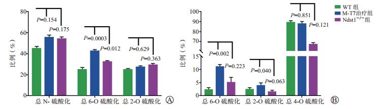
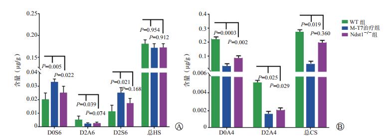
目的 探讨小鼠供肾选择性缺失N-脱乙酰基酶-N-磺基转移酶(Ndst)1对肾移植术后受体小鼠急性排斥反应的影响及其机制。 方法 建立小鼠肾移植模型。根据品系和处理方式不同,将小鼠分为3组:野生型(WT)组(n=10)、Ndst1-/-组(n=9)、黏液瘤病毒T7蛋白(M-T7)治疗组(n=7)。术后第10日对各组小鼠实施安乐死,对各组小鼠移植肾分别进行病理学检查和糖胺聚糖(GAGs)中的双糖[硫酸乙酰肝素(HS)和硫酸软骨素(CS)]含量分析。分析病理学评分与双糖含量变化的相关性。 结果 与WT组比较,Ndst1-/-组和M-T7治疗组小鼠移植肾组织的早期排斥反应的总体病理学评分显著降低(均为P < 0.05)。与WT组比较,Ndst1-/-组小鼠移植肾组织中HS的6-O硫酸化比例、D0S6含量显著增加(均为P < 0.05),CS的D0A4和D2A4含量显著下降(均为P < 0.05)。与WT组比较,M-T7治疗组小鼠移植肾组织中HS的6-O硫酸化比例、CS的6-O硫酸化和2-O硫酸化比例、HS的D0S6和D2S6含量均显著增加(均为P < 0.05),HS的D2A0含量、CS的D0A4和D2A4含量明显降低(均为P < 0.05)。小鼠移植肾组织中双糖含量变化与急性排斥反应相关病理学评分均具有相关性(均为P < 0.05)。 结论 供肾选择性缺失Ndst1可减轻肾移植术后的急性排斥反应,其机制可能与移植肾组织中双糖HS的6-O硫酸化比例增加有关。 Abstract:Objective To investigate the effect and mechanism of selective deletion of N-deacetylase-N-sulfotransferase (Ndst) 1 in donor kidney on the acute rejection after renal transplantation in the recipient mice. Methods A mouse model undergoing renal transplantation was established. The mice were divided into 3 groups according to the strains of mice and treatment: wild type (WT) group (n=10), Ndst1-/- group (n=9) and myxoma virus T7 protein(M-T 7)treatment group (n=7). The mice were euthanized at postoperative 10 d. Pathological examination and the content of disaccharide [heparan sulfate (HS) and chondroitin sulfate (CS)] in glycosaminoglycans (GAGs) were analyzed. The correlation between pathological score and changes of disaccharide content was analyzed. Results Compared with the WT group, the overall pathological scores of early rejection of the transplanted kidney tissues were significantly lower in the Ndst1-/- and M-T7 treatment groups (both P < 0.05). Compared with the WT group, the percentage of 6-O sulfation and the content of D0S6 in HS were significantly increased, whereas the contents of D0A4 and D2A4 in CS were significantly decreased in the transplanted kidney tissues in the Ndst1-/- group (all P < 0.05). Compared with the WT group, the percentage of 6-O sulfation in HS, the percentages of 6-O sulfation and 2-O sulfation in CS and the contents of D0S6 and D2S6 in HS were significantly increased, whereas the content of D2A0 in HS, the contents of D0A4 and D2A4 in CS were significantly decreased in the transplanted kidney tissues in the M-T7 treatment group (all P < 0.05). The changes of disaccharide content in the transplanted kidney tissues of mice were significantly correlated with pathological scores related to acute rejection (all P < 0.05). Conclusions Selective deletion of Ndst1 of the donor kidney can alleviate acute rejection after renal transplantation, and the mechanism may be related to the increased 6-O sulfation ratio of disaccharide HS in the transplanted kidney tissues. -

图 1 图 1各组小鼠移植肾组织的病理学图片及病理学评分
A图为WT组小鼠的移植肾组织(HE,×200); B图为Ndst1-/-组小鼠的移植肾组织(HE,×200); C图为M-T7治疗组小鼠的移植肾组织(HE,×200); D图为各组总体病理学评分比较
Figure 1. Pathological images and pathology scores of transplanted kidney tissues of mice in each group

图 2 各组小鼠移植肾组织中HS以及CS硫酸化指标的比较
A图为各组HS的硫酸化指标,包括总N-硫酸化、总6-O硫酸化和总2-O硫酸化比例; B图为各组CS的硫酸化指标,包括总6-O硫酸化、总2-O硫酸化和总4-O硫酸化比例
Figure 2. Comparison of sulfation indexes of HS and CS in transplanted kidney tissues of mice among each group
-
[1] 谭建明, 王萍.间充质干细胞改善实体器官移植效果的临床研究[J/CD].中华细胞与干细胞杂志(电子版), 2016, 6(5): 263-269. DOI: 10.3877/cma.j.issn.2095-1221.2016.05.001.TAN JM, WANG P. Mesenchymal stem cells to improve solid organ transplant outcome: clinical trials[J/CD]. Chin J Cell Stem Cell(Electr Edit), 2016, 6(5): 263-269. DOI: 10.3877/cma.j.issn.2095-1221.2016.05.001. [2] 王红宇, 焦宪法, 牛杏果, 等.伴急性肾损伤的脑死亡器官捐献供者供肾移植治疗的体会[J].器官移植, 2017, 8(6): 424-429. DOI: 10.3969/j.issn.1674-7445.2017.06.003.WANG HY, JIAO XF, NIU XG, et al. Experience of clinical efficacy of renal transplantation from donors of donation after brain death complicated with acute kidney injury[J]. Organ Transplant, 2017, 8(6): 424-429. DOI: 10.3969/j.issn.1674-7445.2017.06.003. [3] NACIF LS, PINHEIRO RS, PÉCORA RA, et al. Late acute rejection in liver transplant: a systematic review[J]. Arq Bras Cir Dig, 2015, 28(3): 212-215. DOI: 10.1590/S0102-67202015000300017. [4] SHIMIZU T, TANABE T, SHIRAKAWA H, et al. Clinical and pathological analysis of transplant glomerulopathy cases[J]. Clin Transplant, 2012, 26(Suppl 24): 37-42. DOI: 10.1111/j.1399-0012.2012.01639.x. [5] 姚杏红, 曾烨.血管内皮糖萼研究进展[J].四川解剖学杂志, 2013, 21(4): 42-46. DOI: 10.3969/j.issn.1005-1457.2013.089.YAO XH, ZENG Y. The research progress of endothelial glycocalyx[J]. Sichuan J Anat, 2013, 21(4): 42-46. DOI: 10.3969/j.issn.1005-1457.2013.089. [6] GE XN, HA SG, RAO A, et al. Endothelial and leukocyte heparan sulfates regulate the development of allergen-induced airway remodeling in a mouse model[J]. Glycobiology, 2014, 24(8): 715-727. DOI: 10.1093/glycob/cwu035. [7] PROUDFOOT AE, BONVIN P, POWER CA. Targeting chemokines: pathogens can, why can' t we?[J]. Cytokine, 2015, 74(2): 259-267. DOI: 10.1016/j.cyto.2015.02.011. [8] ROPS AL, LOEVEN MA, VAN GEMST JJ, et al. Modulation of heparan sulfate in the glomerular endothelial glycocalyx decreases leukocyte influx during experimental glomerulonephritis[J]. Kidney Int, 2014, 86(5): 932-942. DOI: 10.1038/ki.2014.115. [9] 陈昊, 韩志坚, 彭正奎, 等.应用肾静脉袖套式血管吻合技术建立小鼠原位肾移植模型[J].中华器官移植杂志, 2015, 36(10): 611-615. DOI: 10.3760/cma.j.issn.0254-1785.2015.10.008.CHEN H, HAN ZJ, PENG ZK, et al. Orthotopic kidney transplantation in mice: technique using cuff for renal vein anastomosis[J]. Chin J Organ Transplant, 2015, 36(10): 611-615. DOI: 10.3760/cma.j.issn.0254-1785.2015.10.008. [10] DAI E, LIU LY, WANG H, et al. Inhibition of chemokine-glycosaminoglycan interactions in donor tissue reduces mouse allograft vasculopathy and transplant rejection[J]. PLoS One, 2010, 5(5): e10510. DOI: 10.1371/journal.pone.0010510. [11] BAR Y, BARREGARD L, SALLSTEN G, et al. Quantitative and semi-quantitative histopathological examination of renal biopsies in healthy individuals, and associations with kidney function[J]. APMIS, 2016, 124(5): 393-400. DOI: 10.1111/apm.12520. [12] ABIDI J, AMMAR S, BEN BRAHIM S, et al. Use of ultra-high-performance liquid chromatography coupled with quadrupole-time-of-flight mass spectrometry system as valuable tool for an untargeted metabolomic profiling of Rumex tunetanus flowers and stems and contribution to the antioxidant activity[J]. J Pharm Biomed Anal, 2018, 162: 66-81. DOI: 10.1016/j.jpba.2018.09.001. [13] BUTTIGIEG J, AGIUS-ANASTASI A, SHARMA A, et al. Early urological complications after kidney transplantation: an overview[J]. World J Transplant, 2018, 8(5): 142-149. DOI: 10.5500/wjt.v8.i5.142. [14] LEMOINE M, GUERROT D, BERTRAND D. Focusing on kidney transplantation in the elderly[J]. Nephrol Ther, 2018, 14(2): 71-80. DOI: 10.1016/j.nephro.2017.06.003. [15] RODRÍGUEZ CASTELLANOS FE, DOMÍNGUEZ QUINTANA F, SOTO ABRAHAM V, et al. Classification of acute rejection episodes in kidney transplantation: a proposal based on factor analysis[J]. Iran J Kidney Dis, 2018, 12(2): 123-131. [16] DE MURO P, FAEDDA R, MASALA A, et al. Kidney post-transplant monitoring of urinary glycosaminoglycans/proteoglycans and monokine induced by IFN-γ (MIG)[J]. Clin Exp Med, 2013, 13(1): 59-65. DOI: 10.1007/s10238-012-0178-5. [17] POMIN VH, PARK Y, HUANG R, et al. Exploiting enzyme specificities in digestions of chondroitin sulfates A and C: production of well-defined hexasaccharides[J]. Glycobiology, 2012, 22(6): 826-838. DOI: 10.1093/glycob/cws055. -

 下载:
下载:

 点击查看大图

图(3)
计量
- 文章访问数: 117
- HTML全文浏览量: 60
- PDF下载量: 5
- 被引次数: 0


